

 Book An Appointment



Updated MDI Surgery Technique
Dr. Garcia specializes in complex knee, shoulder and elbow sports surgeries. He has prepared a number of surgical videos below to help patients better understand their procedures. He is frequently updating his surgical video database so check back soon for further updates.
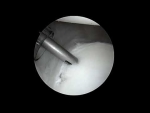
This video demonstrates Dr. Grant Garcia’s updated surgical technique for managing Multidirectional Shoulder Instability (MDI). This condition represents a challenging clinical scenario, typically seen in patients with severe, systemic generalized joint laxity who experience frequent dislocations or sliding (subluxations) in multiple directions (anterior, posterior, and inferior).
In this specific case, the patient suffered from hundreds of instability events and a circumferential, 360-degree labral tear. Dr. Garcia utilizes an all-knotless fixation technique with small 1.8 mm anchors, which minimizes hardware irritation and ensures a smooth, low-profile reconstruction.
The procedure focuses on reducing joint volume and tightening the loose capsule through the following surgical steps:
- Mobilization & Elevation: Dr. Garcia begins by arthroscopically debriding the frayed tissue and carefully elevating the torn labrum away from the shoulder socket (glenoid) rim. This allows him to perform a comprehensive, simultaneous repair of both the labrum and the stretched-out joint capsule.
- Posterior & Inferior Capsular Reefing: Starting low on the back side of the joint, the first knotless FiberTack anchor is inserted. Dr. Garcia utilizes a specialized "pinch and roll" technique with a rotational suture lasso, pulling a substantial pleat of the lax capsule inward. The suture is passed and automatically retrieved within the cannula to speed up the process. Cinching it down re-tensions the inferior glenohumeral ligament (IGHL)—the primary lower hammock holding the shoulder bone in place.
- Circumferential Stabilization: Moving systematically around the socket, a second and third knotless anchor are deployed to fix the superior and posterior labral boundaries. Dr. Garcia takes great care to keep the small suture lines clear of tangles to protect the fine locking mechanisms of the all-knotless constructs.
- Anterior Plication: Shifting focus to the front of the shoulder, low inferior anchors are placed. The same "pinch and roll" technique is applied to gather the redundant anterior capsule, building a robust anterior tissue bumper. The sutures are placed and temporarily saved, allowing Dr. Garcia to perform a sequential, balanced tensioning from bottom to top (inferior to superior) to evenly tighten the entire joint space.
- Closing the Posterior Capsular Rent: To address any remaining posterior redundancy, Dr. Garcia uses the rotational lasso to pass a thick suture tape across the posterior capsule, retrieves it with an arthroscopic BirdBeak passer, and ties it down using alternating half-hitches.
The final visualization shows a highly compact, balanced, and tightly plicated shoulder capsule in all directions. By reshaping the native capsule and securing the 360-degree labrum, the redundant joint volume is eliminated, restoring global structural stability to a highly unstable shoulder joint.







