

 Book An Appointment



Updated MCL reconstruction with internal brace
Dr. Garcia specializes in complex knee, shoulder and elbow sports surgeries. He has prepared a number of surgical videos below to help patients better understand their procedures. He is frequently updating his surgical video database so check back soon for further updates.
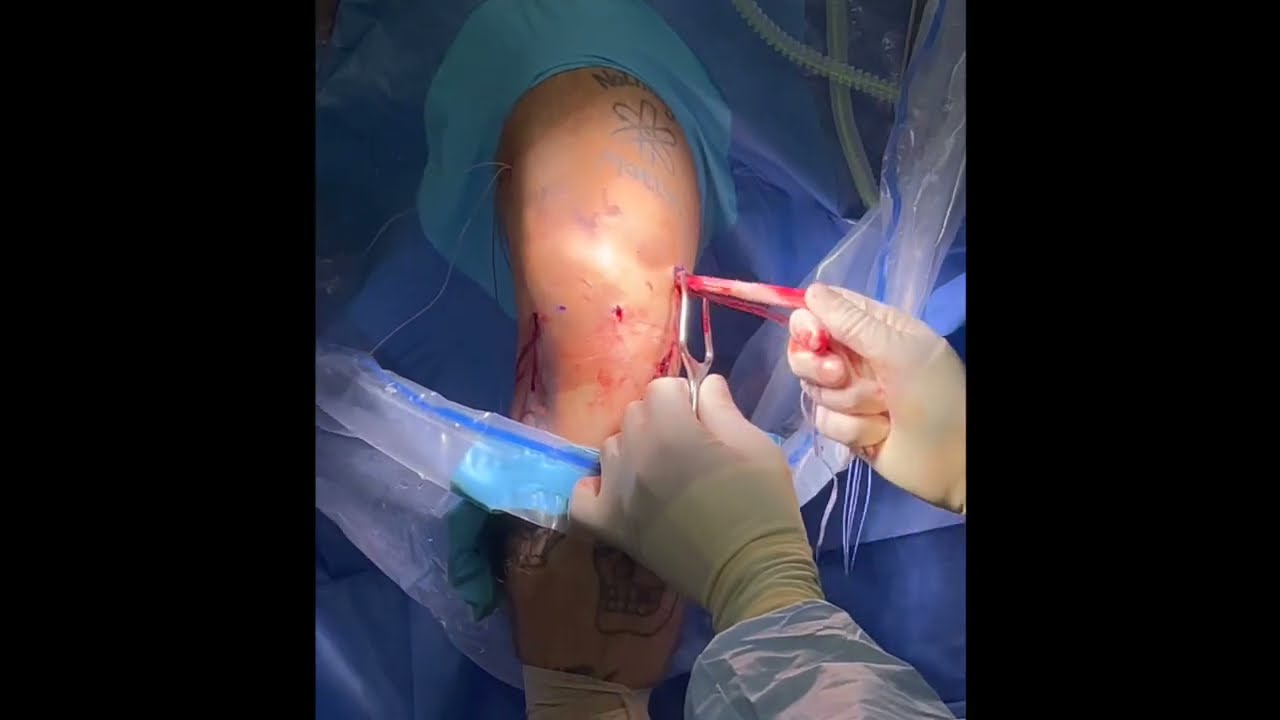

This video features Dr. Garcia demonstrating an updated Medial Collateral Ligament (MCL) reconstruction technique using an Achilles allograft, tightrope fixation, and an internal brace. The patient had a chronic MCL tear that may have developed following a previous tibial plateau fracture.
The surgical procedure involves the following steps:
- Graft Preparation and Femoral Fixation: The surgeon prepares the Achilles allograft by removing the bone block. They attach a FiberTag tightrope system to it, pull the fixation button through to the lateral cortex, and confirm proper positioning with an X-ray. The graft is sequentially tightened, dunking roughly 20 mm of it into a pre-drilled femoral socket to avoid bottoming out.
- Isometry and Deep Repair: After identifying the point of isometry (the position where the ligament maintains steady tension during knee movement), a 2.6 mm FiberTack anchor with tapes is placed to reconstruct the deep layer of the MCL.
- Tibial Fixation and Tensioning: The surgeon whip-stitches both sides of the graft for a stronger two-part fixation. The leg is bent to approximately 20 degrees with applied resistive force while the tibial anchors are placed. The surgeon carefully balances tension, sequentially tightening the structural sutures while avoiding overtightening the internal brace tapes to prevent over-constraining the joint.
- Final Adjustments: The tightrope system allows the surgeon to pull out any mechanical creep and retension the graft after the tibial fixation is complete. The deep sutures are tied down to provide robust compressive force.
- Verification: The reconstruction achieves optimal structural tension with zero gapping, and the surgeon performs a final arthroscopic evaluation inside the joint to confirm the knee moves smoothly without being over-constrained.







