

 Book An Appointment



Updated Distal Tibial Allograft Glenoid Reconstruction Surgery
Dr. Garcia specializes in complex knee, shoulder and elbow sports surgeries. He has prepared a number of surgical videos below to help patients better understand their procedures. He is frequently updating his surgical video database so check back soon for further updates.
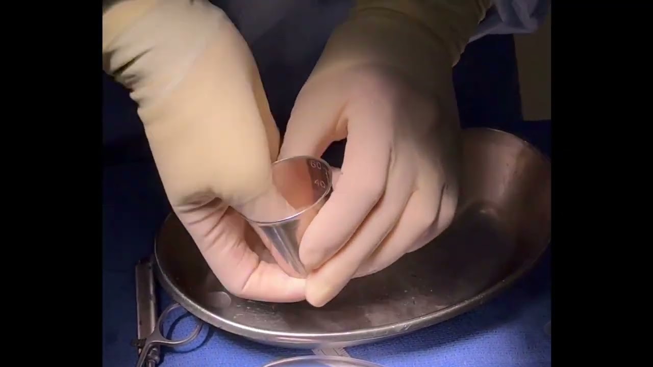

This video features Dr. Grant Garcia demonstrating his specialized surgical technique for an open Distal Tibial Allograft (DTA) glenoid reconstruction. This advanced bony reconstruction procedure is indicated for patients with critical anterior glenoid bone loss—in this case, a severe 25% bone loss defect—often resulting from recurrent, high-energy anterior shoulder dislocations or a failed previous stabilization surgery.
While a Latarjet procedure uses a local coracoid autograft, the distal tibial allograft is an excellent alternative because its natural articular geometry perfectly matches the native curvature of the glenoid socket, effectively restoring joint volume and structural stability without donor-site morbidity.
The procedure is executed through the following key phases:
- Graft Selection and Pre-Planning: Using an specialized guide system (Arthrex), Dr. Garcia measures the defect. He aims for a 7 mm distal tibial allograft cut at a precise 10-degree angle. He notes that matching the native anatomy perfectly is crucial; over-stuffing the joint space can actually pathologically increase bone resorption over time.
- Biologic Graft Optimization: On the back table, the graft is carefully cut, marked for spatial orientation, and pulsed-lavaged. Dr. Garcia uses carbon dioxide (CO2) to meticulously clear out the marrow elements from the bone graft. This process dramatically enhances its porosity, allowing it to act like a sponge when it is soaked in the patient's pre-spun Platelet-Rich Plasma (PRP) to accelerate bone-to-bone integration.
- Managing Failed Hardware & Surface Prep: Shifting to the open surgical site, Dr. Garcia exposes the front of the joint. He removes multiple loose or unabsorbed anchors from a failed prior surgery and aggressively rasps the anterior glenoid neck to form a flat, bleeding bone bed.
- Parallel Guide Pin Placement: Utilizing a parallel wire guide system, parallel guide pins are drilled directly into the glenoid defect. The lengths are measured and verified against pre-operative CT scans.
- Fixation with a Two-Hole Compression Plate: The PRP-soaked distal tibial graft is slotted over the guide wires. Dr. Garcia uses a low-profile two-hole compression plate rather than standalone screws. This plate distributes compression forces evenly across the graft bone without introducing localized stress fractures. Fully threaded, cannulated screws are advanced into the plate.
- Soft-Tissue Integration: Before final torque is applied to the screws, Dr. Garcia slides two heavy FiberWire sutures underneath the plate. These are reserved to tie down and plicate the surrounding capsule/labrum later on to form a soft-tissue barrier.
- Final Hardware Tensioning: The cannulated screws are hard-tightened to fully seat the graft. Because distal tibial bone is highly dense, the surgeon can apply robust compression without risking structural fragmentation.
The final intraoperative inspection shows a perfectly flush bony reconstruction with absolutely no step-off or overhang, restoring the native structural arc of the shoulder socket.







