

 Book An Appointment



Posterior Lateral Corner Reconstruction
Dr. Garcia specializes in complex knee, shoulder and elbow sports surgeries. He has prepared a number of surgical videos below to help patients better understand their procedures. He is frequently updating his surgical video database so check back soon for further updates.
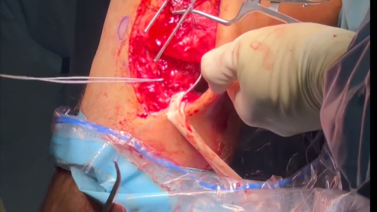

This video, titled "Posterior Lateral Corner Reconstruction," demonstrates an anatomic reconstruction of the posterolateral corner (PLC) of the knee using a semitendinosus allograft (donor tissue). The procedure is performed on a patient who previously underwent ACL surgery but requires a PLC reconstruction due to severe, chronic lateral knee instability.
This is a particularly complex case because the anatomy is severely scarred down, and the patient has a complete avulsion injury where both the biceps femoris tendon and the Lateral Collateral Ligament (LCL) have torn completely off the head of the fibula bone.
The core surgical phases demonstrated in the video include:
- Surgical Exposure and Critical Nerve Isolation: The surgeon makes a lateral incision. Because the tissue is heavily scarred, a fascial sheet is carefully removed to visualize the landmarks. Crucially, the peroneal nerve is dissected out first and isolated. Isolating this major nerve ensures it can be physically protected from drills and sharp instruments throughout the operation.
- Managing the Avulsed Tissue: The torn biceps femoris and LCL are identified as a single detached sheet of tissue. The surgeon safely preserves this layer to repair it back to the bone later in the procedure.
- Drilling the Femoral Tunnels: To recreate the LCL and popliteofibular ligament (PFL) femoral attachments, the surgeon identifies the lateral epicondyle and its surrounding sulcus. A small vertical arthrotomy is made into the joint capsule to expose the target zones. Two separate, precisely measured tunnels are drilled into the femur (about 35mm deep) based on the thickness of the allograft.
- Drilling the Fibular Tunnel: Using a specialized centering guide, a tunnel is mapped out straight through the center of the fibular head from front to back (anterior to posterior). The surgeon places a spoon-shaped metal protective device behind the bone to shield the isolated peroneal nerve and deep tissues while the drill reams out a 20-30mm passage.
- Graft Passage and Femoral Fixation: The semitendinosus allograft is threaded into the knee tunnels. The surgeon first secures the LCL portion of the graft inside the femoral tunnel using a rigid PEEK interference screw. The graft is then routed down through the new fibular head tunnel and directed back up toward the second femoral socket to recreate the popliteofibular ligament.
- Biceps Femoris Repair and Final Tensioning: To fix the completely detached biceps femoris muscle, the surgeon weaves heavy-duty suture down and back through the tendon using a locking Krakow stitch. With the patient's knee flexed to 45 degrees, the graft limbs and the biceps femoris sutures are drawn tight. A 6mm SwivelLock anchor is driven directly into the fibular head to secure both the soft-tissue reconstruction and the reattached muscle simultaneously.
- Verification: The surgeon cycles the leg to ensure the graph handles regular stress without bottoming out. The joint is examined arthroscopically to visually verify that the abnormal outer knee gapping and laxity have been completely eliminated.







