

 Book An Appointment



Midsubstance Patella Tendon Repair with Hamstring Augmentation
Dr. Garcia specializes in complex knee, shoulder and elbow sports surgeries. He has prepared a number of surgical videos below to help patients better understand their procedures. He is frequently updating his surgical video database so check back soon for further updates.
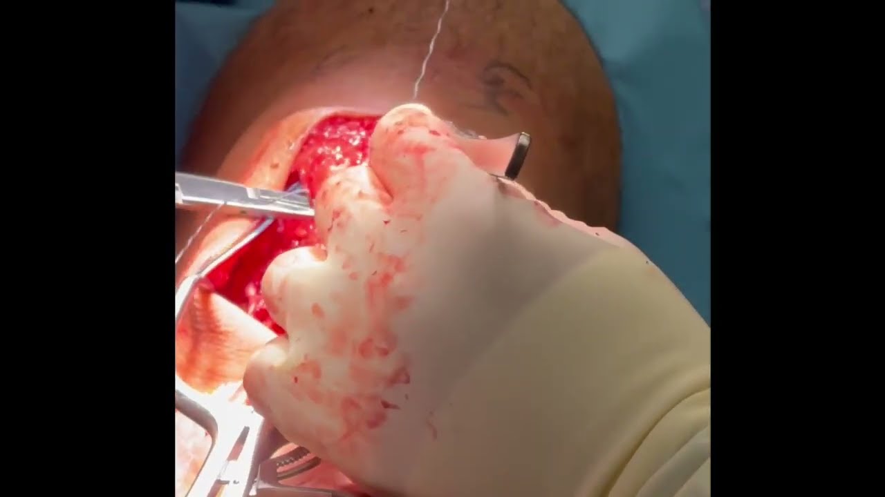
In this video, Dr. Grant H. Garcia demonstrates his surgical technique for managing a midsubstance patellar tendon rupture. Because the rupture occurs within the tissue itself rather than tearing directly off the bone, this type of injury is quite uncommon and technically challenging to repair due to the poor quality of tendon available on either side. To ensure structural integrity, Dr. Garcia augments the repair with a harvested hamstring autograft and an internal brace.
The procedure is executed through several critical stages:
- Anchor Placement & Reapproximation: Two knee FiberTack anchors are placed in the patella and one is driven into the tibia. Dr. Garcia performs a running Krackow stitch between the torn segments of the tendon. Using a minor tension-slide technique with the knee held in full extension, the two ends of the midsubstance tear are pulled together and reapproximated tightly. The native patellar sutures are isolated to serve as part of the future internal brace.
- Preventing Patella Baja: The sutures are sequentially tied across the midsubstance gap. Dr. Garcia emphasizes the importance of balancing the tension during this phase—over-tightening the repair will pull the kneecap too low, creating an unwanted condition known as patella baja. A final suture from the tibia is paired to the upper tendon segment to reduce tissue creep and potential separation.
- Crisscross Internal Bracing: To protect the repair during aggressive post-operative range of motion, a dual-limb internal brace is created in a crisscross fashion using limbs from the FiberTack anchors. Crucially, this internal brace is tensioned with the knee bent to approximately 70 degrees; tensioning it in full extension would dangerously over-constrain the construct.
- Hamstring Autograft Augmentation: The hamstring tendon is harvested but intentionally left attached at its native tibial insertion. The semitendinosus (referred to as the semimembranosus in the narrative tracking) and gracilis tendons are draped over the repair in a crisscross pattern to add vascularity and bulk.
- Final Anchor & Motion Check: With the knee brought back into full extension, the distal end of the longer tendon graft is measured, truncated, and dunked into a bone tunnel with a final anchor. The graft limbs are then sutured directly down to the surrounding construct.
A final range of motion check shows no gapping at the repair site. The internal brace is designed to remain slack during initial movement and only fully engages past 70 degrees of flexion to act as a definitive check-rein against overload.







