

 Book An Appointment



Live MACI Arthro: Rods & Cones System
Dr. Garcia specializes in complex knee, shoulder and elbow sports surgeries. He has prepared a number of surgical videos below to help patients better understand their procedures. He is frequently updating his surgical video database so check back soon for further updates.
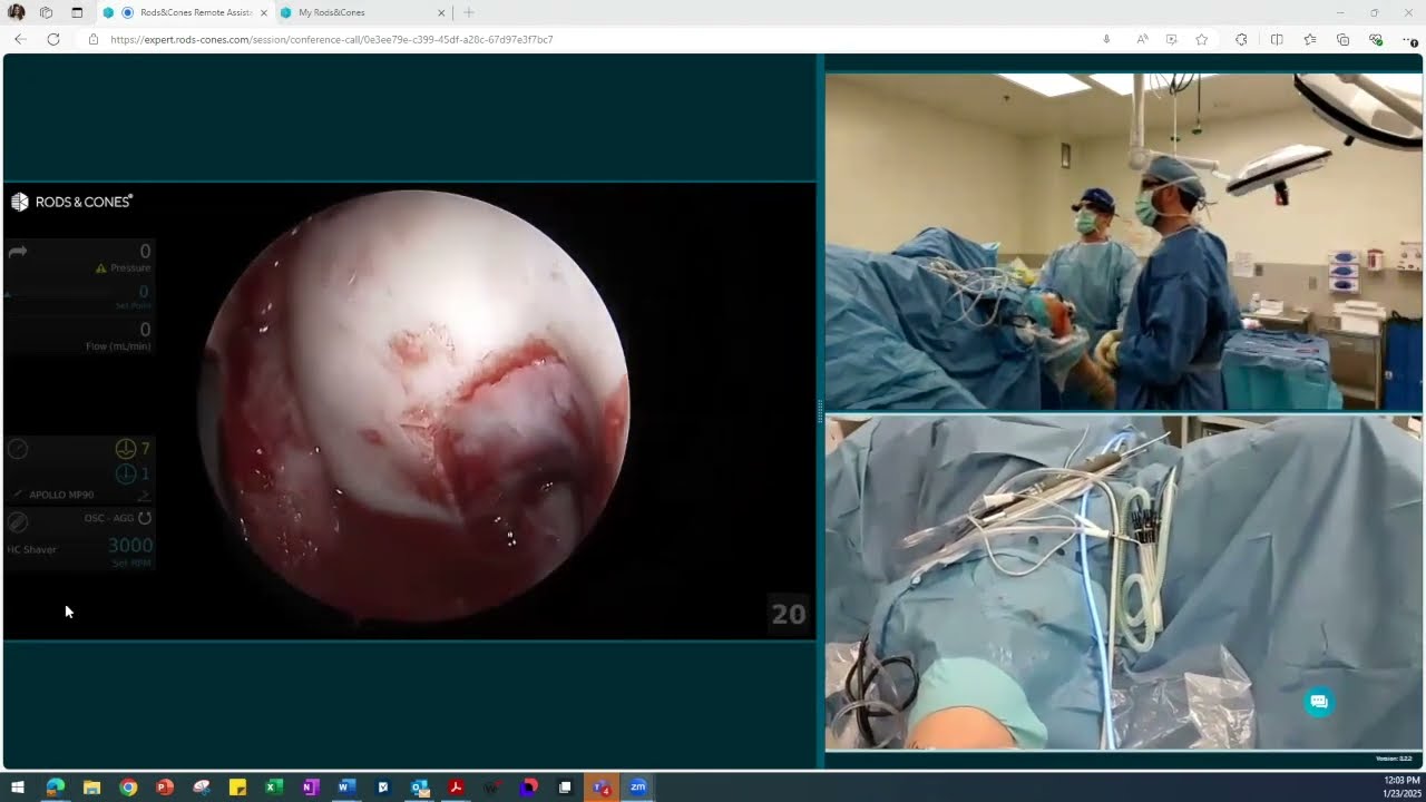
This video, presented by Dr. Grant H. Garcia, features a live surgical demonstration of a MACI Arthro procedure to repair a trochlear defect (a cartilage defect in the groove of the knee). The surgery utilizes the Rods & Cones video system, providing simultaneous views of both the internal arthroscopic camera and the external field in the operating room.
Surgical Breakdown:
- Site Preparation & Visualization: The surgeon begins by cleaning up the fat pad and internal scarring to maximize visibility. Working through standard arthroscopic portals, the team debrides (cleans out) the loose or damaged cartilage surrounding the defect.
- Cannula Placement & Precision Cutting: With the knee held in a flexed position, a small 2 cm incision is made to introduce a 15 mm MACI Arthro cannula. A specialized cutter tool outlines the margins of the injury, and curettes are carefully used to strip away soft cartilage down to a hard, stable edge. The surgeon emphasizes that keeping the border precise—leaving less than a 1 to 2 mm gap around the implant—is crucial for a successful fit.
- Hemostasis (Controlling Bleeding): Once the bone is prepped and the surgical tourniquet is released, the team addresses capillary bleeding in the defect bed. They apply epinephrine pads, gel thrombin, and Tisseel fibrin sealant, using a gauze-covered finger to firmly compress the area and create a clean, dry surface.
- Loading the MACI Membrane: After performing a trajectory "dry run" with a delivery tool called a V-Shuttle, the surgeon prepares the MACI implant. The biological membrane is carefully oriented with the active cell-side facing properly and loaded onto the protective arms of the V-Shuttle.
- Implantation & Smoothing: A light layer of Tisseel glue is spread onto the prepared bone, and the V-Shuttle deploys the membrane directly into the knee (cell-side down, shiny side up). The surgeon utilizes a small plastic tip to gently maneuver the membrane flat into place, ensuring an excellent fill and smoothing out any trapped air bubbles.
- Final Seal & Stability Check: Starting from the top to let gravity assist, the surgeon applies a heavy layer of Tisseel sealant around the graft edges, letting it dry for three minutes between rounds to secure it against joint fluid leakage. To conclude the procedure, the knee is cycled through a 0-to-90-degree range of motion to visually confirm that the MACI membrane remains perfectly stable and securely adhered to the bone.







