

 Book An Appointment



Dr.Garcia’s newest technique for arthroscopic glenoid fracture repair
Dr. Garcia specializes in complex knee, shoulder and elbow sports surgeries. He has prepared a number of surgical videos below to help patients better understand their procedures. He is frequently updating his surgical video database so check back soon for further updates.
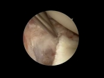

This video features Dr. Grant Garcia demonstrating an advanced arthroscopic technique for internal fixation of a glenoid fracture (bony Bankart lesion). The patient presented with an acute injury causing an 18% bone loss deficit along the front edge of the shoulder socket, which requires direct reconstruction to prevent recurrent dislocations.
The procedure focuses on restoring the native bone volume and rebuilding the labral bumper through the following key clinical phases:
- Site Preparation & Portal Strategy: Dr. Garcia begins by using an arthroscopic rasp to clean out the fracture site, debriding loose debris while carefully preserving the detached bone fragment and its connection to the native labrum. To establish an optimal angle for fixation, he creates a specialized 5 o'clock subscapularis working portal.
- Securing the Bone Fragment: A first anchor (SutureTack) is implanted low and centrally within the defect. Dr. Garcia uses a specialized rotational lasso tool to efficiently pass suture tapes through the capsule and around the bony piece. This tool allows him to capture and retrieve the sutures automatically without having to shuttle them outside the cannula, preventing the fragment from separating from the labrum.
- Suspended Knotless Fixation: To hold the bone fragment in a reduced position, a knotless FiberTack anchor is inserted inferiorly. The rotational lasso is used to take a secure bite of the inferior capsule (taking care not to dive too deep into the axilla). Pulling the tension on this anchor dynamically draws the bony piece back into its proper anatomical position.
- Double-Row Bridging Construct: A superior anchor is placed just above the fracture zone. Dr. Garcia passes a horizontal mattress stitch across the tissue to create a double-row construct. This prevents the fragment from rotating or over-reducing. The suture limbs from the initial fixation are then spanned over the top of the bone piece and locked down with a PushLock anchor, providing rigid, compression-based internal fixation.
- Final Labral Re-tensioning: After sequentially retensioning and cutting the inferior and superior anchor tails to finalize bone compression, Dr. Garcia identifies a slight residual laxity in the soft tissue. He places one final FiberTack anchor to cinch down the remaining labrum, fully recreating the thick anterior tissue bumper.
The final inspection demonstrates that the 18% glenoid bone deficit has been completely restored to its original shape and the labral barrier is fully tensioned to eliminate future joint instability.







