

 Book An Appointment



Dr. Garcia’s New Tensionable Tibial Spine Technique
Dr. Garcia specializes in complex knee, shoulder and elbow sports surgeries. He has prepared a number of surgical videos below to help patients better understand their procedures. He is frequently updating his surgical video database so check back soon for further updates.
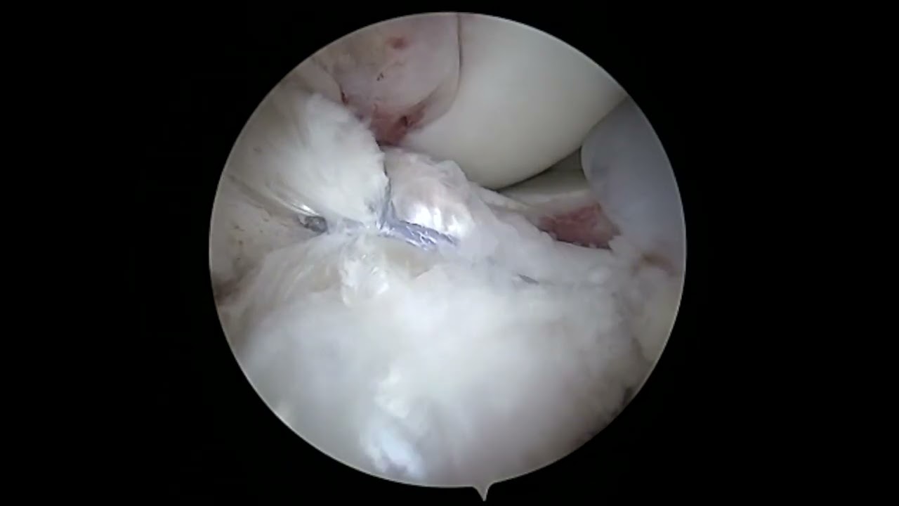

This video, presented by orthopedic surgeon Dr. Grant H. Garcia, demonstrates an updated technique for treating an ACL tibial spine avulsion fracture using a two-tunnel tensionable TightRope fixation.
An avulsion fracture occurs when the ACL bone attachment site pulls away from the tibia rather than the ligament tearing. This two-tunnel layout adds high biomechanical strength, allowing the patient to start immediate weight bearing after surgery.
Surgical Breakdown:
- Site Preparation: The large avulsion piece—including a part of the lateral meniscus root—is evaluated. The surgeon cleans up the fracture site using a radiofrequency device and a shaver, prepping it to ensure optimal bone-to-bone healing. Passport cannulas are placed in the medial and lateral portals to facilitate clean suture passing.
- Tunnel Drilling: A specialized guide is used to place two 2.4 mm cannulated pins through the tibia, spaced roughly 1 centimeter apart. The pins are slightly backed out from the joint space to protect the native tissues during prep.
- Passing the FiberRings: The surgeon uses a Knee Scorpion to pass a 35 mm FiberRing suture device directly through the bundles of the native ACL. This functions essentially like an ACL repair but in reverse. Two distinct passing stitches (one loop side, one tail side) are established.
- TightRope Engagement & Reduction: The passing sutures are pulled up through the tibial tunnels and out the cannulas. The FiberRings are interlocked with the internal repair device, and everything is pulled back down into the bone tunnels. This creates a secure crossover link over the bone fragment.
- Compression and Final Cinching: As the sutures are pulled tight, the bone fragment is compressed back down. The surgeon targets a slight "over-reduction" (pulling the fragment just past flat) and trims the very front edge to eliminate any potential anterior impingement when the patient extends their knee.
- Dual Button Security: The sutures are threaded through two cortical buttons on the outside of the shin bone. With the knee held in full extension, the TightRope loops are sequentially tensioned, drawing the bone fragment flush into its socket.
- Fixation Backup: After checking the final reduction on an intraoperative X-ray, the suture strands from both buttons are tied down. To provide ultimate security against loosening, the remaining strands are loaded and sunk directly into a final SwiveLock anchor. This stable, rigid design avoids the risk of post-operative knee stiffness and permits rapid restoration of movement and weight bearing.







