

 Book An Appointment



Dr. Garcia’s new technique for elbow UCL reconstruction
Dr. Garcia specializes in complex knee, shoulder and elbow sports surgeries. He has prepared a number of surgical videos below to help patients better understand their procedures. He is frequently updating his surgical video database so check back soon for further updates.
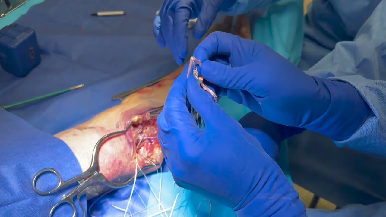
This video features Dr. Grant H. Garcia demonstrating his new technique for an elbow ulnar collateral ligament (UCL) reconstruction utilizing knotless fixation. This advanced method replicates a stable, native-like double-bundle pattern to treat elbow instability.
Here is a summary of the procedure shown in the video:
- Exposure and Nerve Protection: The surgeon initiates the medial approach, carefully exposing the joint. The ulnar nerve is safely isolated and tagged more proximally to keep it protected and out of the way for a later transposition. The native, damaged UCL tissue is prepared for the reconstruction.
- Distal Fixation on the Ulna: The surgeon identifies the sublime tubercle and places a guide wire at least 5 millimeters away from the joint line. A 5mm reamer is used to drill a bone socket to a precise depth of 15 millimeters. A palmaris longus (PL) tendon autograft is loaded onto a 4.75mm fork-tip SwiveLock anchor along with a suture tape that will serve as an internal brace.
- Recreating the Double Bundle: The surgeon holds both strands of the tendon taut to create a double-bundle technique. The anchor is securely engaged into the ulnar socket. Upon testing, the construct shows excellent structural fixation with the tendon strands exiting neatly on both sides.
- Humeral Socket Preparation: With the patient's arm resting at 30 degrees of flexion, the surgeon marks the anatomical insertion footprint on the humeral side. The native tissue at the footprint is removed to create a raw healing bed. The surgeon positions a guide pin more toward the epicondyle rather than too close to the joint line. A bone tunnel is reamed to a depth of roughly 18 millimeters using a 5mm reamer.
- Graft Tensioning and Insertion: To ensure the graft doesn't bottom out in the socket, the entry point is carefully marked. Both limbs of the tendon graft are whip-stitched across 12 to 13 millimeters of tissue, and any excess graft length is trimmed. The two whip-stitched tendon strands, along with the underlying native tissue, are bundled and driven down into the humeral bone tunnel using a standard 4.75mm SwiveLock anchor, creating a tight press-fit.
- Construct Testing and Reinforcement: The elbow is evaluated across its range of motion. It exhibits robust tension at 30 degrees and displays zero restriction or over-constraint in full extension. The surgeon ties alternating half-hitches with the suture tapes over the top to finish the structural internal brace.
- Capsular Closure: To prevent the synthetic tape from experiencing a "windshield wiper" effect against the bone and to fully combine the reconstructive components, the joint capsule is completely repaired and closed over the construct.
The procedure concludes with a valgus stress test, confirming there is absolutely no joint gapping, leaving a highly secure and biomechanically stable reconstruction.







