

 Book An Appointment



Dr. Garcia’s Hay Barrel Technique for Discoid Meniscus Repair
Dr. Garcia specializes in complex knee, shoulder and elbow sports surgeries. He has prepared a number of surgical videos below to help patients better understand their procedures. He is frequently updating his surgical video database so check back soon for further updates.
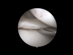
In this video, Dr. Grant H. Garcia demonstrates the "Hay Barrel" technique used for repairing a horizontal tear in a discoid lateral meniscus.
The procedure follows these major surgical steps:
- Debridement & Preparation: After removing the damaged, torn center of the discoid meniscus, a torpedo shaver is used to clean up the edges, aiming for a stable 6 to 8 mm rim. Because there is no root instability, a capsular repair is not required. Dr. Garcia performs trephination to stimulate local blood flow and utilizes a ball rasp to irritate the center of the horizontal tear to aid healing.
- Suture Passing & Compression: A cannula is placed to keep workspace access clear. A specialized suturing device passes a size 0 suture around the meniscus, compressing the horizontal split.
- Sequential Knot Tying: The key to this technique is sequential knot tying. Dr. Garcia notes the importance of keeping the suture posts on the inferior (bottom) surface of the meniscus to keep the knots completely out of the joint space. He alternates angles, passing sutures from both the lateral and medial portals to work cleanly around the tissue.
- Anterior Transition & Final Fixation: Because the anterior midbody can be difficult to access with all-inside devices, Dr. Garcia transitions to using a few inside-out stitches to securely compress and pull the anterior portion back toward the joint capsule.
The final arthroscopic view shows excellent, uniform compression of the horizontal split. Following the procedure, the patient transitions to a non-weightbearing rehabilitation protocol to protect the repair.







