

 Book An Appointment



Dr. Garcia demonstrates his updated MACI Arthro Live Surgery
Dr. Garcia specializes in complex knee, shoulder and elbow sports surgeries. He has prepared a number of surgical videos below to help patients better understand their procedures. He is frequently updating his surgical video database so check back soon for further updates.
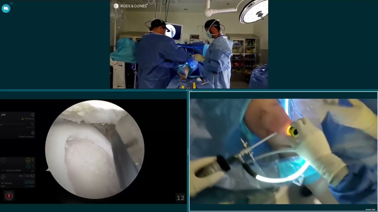

In this surgical video, Dr. Grant H. Garcia demonstrates an arthroscopic MACI (Matrix-induced Autologous Chondrocyte Implantation) procedure on a trochlear defect using the Rods & Cones system. The footage provides a simultaneous first-person operating room view and an arthroscopic view.
The procedure is broken down into the following operational stages:
- Portal Establishment & Visualization: Initial fat pad debridement is performed from both the medial and lateral portals to ensure clear visualization, which is especially critical when switching to a dry arthroscopic environment. Using a spinal needle to identify the correct trajectory perpendicular to the lateral trochlear defect, Dr. Garcia establishes a transpatellar portal. This incision is made while the knee is flexed to safeguard the patellar tendon, then dilated with a 3 cm soft-tissue retracting cannula.
- Defect Preparation: With the knee in near-full extension, a specialized cartilage cutter prepares the borders of the defect. Because accessing the extreme superior and inferior edges can be challenging arthroscopically, a combination of ring curettes, a square tool for flush margins, a shaver, and a small ENT curette are used to meticulously clean the base down to stable walls.
- Hemostasis: The tourniquet is dropped, and fluid is evacuated from the joint. To manage focal bleeding at the bony bed, a Fraser suction tip is introduced, followed by an epinephrine-soaked patty and fibrin sealant (Tisseel). Once absolute hemostasis is achieved, the joint is dried completely, and the tourniquet is inflated again.
- Graft Cutting & Loading: The MACI implant is placed cell-side up on the back table. A custom graft cutter—perfectly matched to the size of the defect preparation tool—is malleted over the implant to cut the precise shape. The graft is loaded equidistant onto a V-Shuttle delivery tool, which utilizes a suction seal to hold the matrix securely.
- Implantation & Fixation: Working in the dry environment, a layer of fibrin sealant is applied to the defect bed. The V-Shuttle is introduced through the central portal and positioned as high as possible on the defect before being deployed. The matrix is gently tapped into place using small touches with a specialized swab. A final layer of fibrin sealant is applied and left to set.
- Stability Assessment: The knee is taken through a full range of motion. The tracking of the patella over the trochlea naturally compresses and contours the graft smoothly into the defect with no displacement or lifting of the edges. The cannula is then removed to complete the procedure.







