

 Book An Appointment



Dr. Garcia demonstrates his technique for subpectoral mini-open biceps tenodesis.
Dr. Garcia specializes in complex knee, shoulder and elbow sports surgeries. He has prepared a number of surgical videos below to help patients better understand their procedures. He is frequently updating his surgical video database so check back soon for further updates.
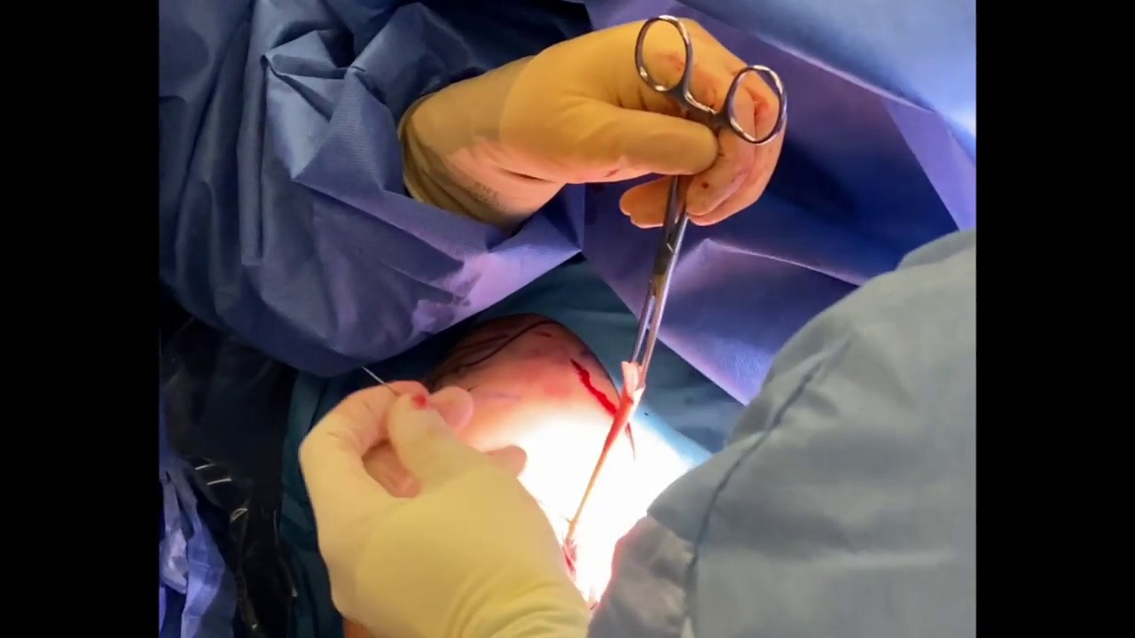

In this video, Dr. Grant Garcia demonstrates his precise surgical technique for an subpectoral open biceps tenodesis. This procedure is commonly performed alongside a rotator cuff repair to address severe biceps tendonitis or tearing by cutting the tendon away from its original attachment inside the shoulder joint and anchoring it lower down on the arm bone (humerus).
The video highlights the following steps of the operation:
- Incision for Improved Cosmesis: Dr. Garcia makes a horizontal incision near the axillary fold (armpit recess) following Langer's lines (the natural lines of skin tension). This specific alignment provides excellent visualization of the lower bicipital groove while yielding a much less noticeable surgical scar compared to traditional vertical incisions.
- Tendon Harvest and Whipping: The diseased portion of the long head of the biceps tendon is pulled out and evaluated. Starting at the musculotendinous junction (where muscle meets tendon), the surgeon weaves a heavy suture tape loop up and down through the tissue, locking it securely at the end. The remainder of the frayed, diseased top part of the tendon is then trimmed away.
- Retractor Placement: To expose the bicipital groove underneath the pectoralis major muscle, retractors are carefully placed. A lateral retractor holds the pectoralis tendon outward, and a small medial "baby Bennett" retractor exposes the lower bony floor of the groove.
- Socket Reaming: A guide wire is drilled centrally directly into the bicipital groove floor. Dr. Garcia then uses a surgical reamer (typically a 6.5 mm size) over the guide wire to create a bone socket designed to house the tendon.
- Interference Screw Fixation: While keeping the patient's arm in a slight amount of flexion to maintain anatomically correct resting muscle tension, a single suture limb is passed through the tip of a 6.25 mm Arthrex Bio-Tenodesis screw. The screw and the whipped tendon are driven together straight into the bone tunnel.
This creates a rigid interference fit, compressing the tendon tightly against the walls of the bone socket. Dr. Garcia notes that recent biomechanical studies favor this interference screw technique because it provides the strongest possible construct for reliable, long-term healing.







