

 Book An Appointment



Dr. Garcia demonstrates his technique for lateral elbow ligament reconstruction for chronic elbow dislocations.
Dr. Garcia specializes in complex knee, shoulder and elbow sports surgeries. He has prepared a number of surgical videos below to help patients better understand their procedures. He is frequently updating his surgical video database so check back soon for further updates.
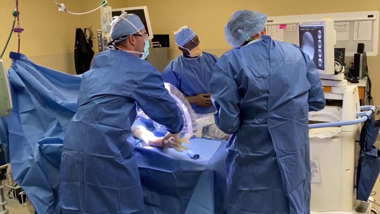
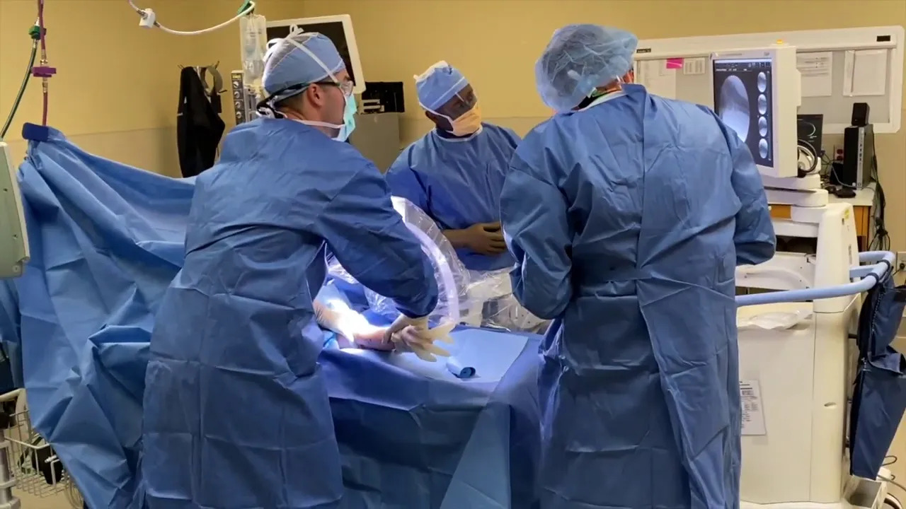
This video demonstrates Dr. Grant H. Garcia's surgical technique for a lateral ulnar collateral ligament (LUCL) reconstruction. This procedure is designed to treat posterolateral rotatory instability (PLRI) resulting from chronic elbow dislocations.
Here is a summary of the procedure shown in the video:
- Pre-operative Confirmation & Exposure: The procedure begins with fluoroscopy (live X-ray) to identify and confirm the instability across all planes. A Kocher approach is then utilized to split the anconeus and extensor tendons, revealing a very thinned and diseased LUCL. A Cobb elevator is used to elevate muscle off the bone down to the ulnar attachment.
- Tunnel Preparation: An arthrotomy is performed through the damaged LUCL tissue. An anterior Hohmann retractor allows visualization of the insertion points. Using fluoroscopy to ensure correct column placement, the surgeon drills a 15-millimeter tunnel into the capitellum (humerus). Turning to the ulna, a matching 15-millimeter tunnel is drilled at the anatomical footprint of the LUCL to allow for a strong single-tunnel fixation.
- Ulnar Graft Fixation: An allograft tendon is secured into the ulnar tunnel using a 4.75 SwiveLock anchor. Maintaining precise tension while inserting the anchor is critical to achieving a solid press-fit and interference fit.
- Capsular Closure & Reinforcement: With the arm positioned at 45 degrees, the surgeon repairs the native diseased LUCL tissue and closes the joint capsule. This creates a solid structural barrier for the new tendon graft to rest upon, which is later sutured directly to the repaired native tissue.
- Humeral Graft Fixation: The proximal end of the tendon graft is then tensioned appropriately with the arm kept at 45 degrees. It is secured into the previously drilled capitellar tunnel using a second 4.75 fork-tip SwiveLock anchor.
- Secondary Internal Brace: The surgeon carefully checks the range of motion to confirm the elbow is neither over-tensioned in full extension nor over-constrained in flexion. Finally, the remaining suture tapes from the anchors are tied together over the graft. This creates a secondary internal brace that provides immediate biomechanical strength to protect the reconstruction.







