

 Book An Appointment



Dr. Garcia demonstrates his technique for arthroscopic rotator cuff repair.
Dr. Garcia specializes in complex knee, shoulder and elbow sports surgeries. He has prepared a number of surgical videos below to help patients better understand their procedures. He is frequently updating his surgical video database so check back soon for further updates.
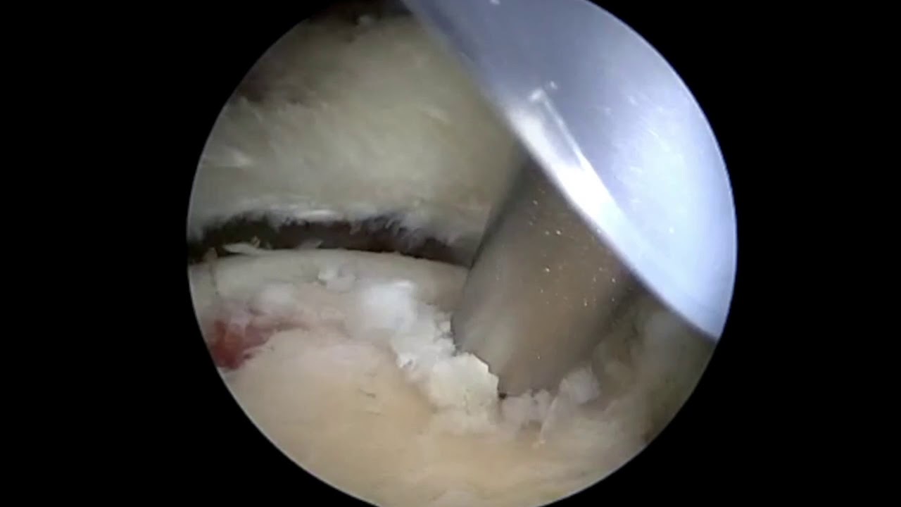

In this video, Dr. Grant Garcia demonstrates his technique for managing a complex, layered arthroscopic rotator cuff repair. The patient initially presented with multiple pathologies, including a comprehensive SLAP (Superior Labrum Anterior to Posterior) tear that required standard debridement, and a heavily diseased long head of the biceps tendon, which was subsequently managed with an open subpectoral biceps tenodesis.
The primary focus of the operation details treating a large, U-shaped central supraspinatus tear that is delaminated (split into two distinct layers of tissue). Recent clinical data demonstrates that separately managing and repairing both layers yields much better long-term structural outcomes.
The procedure is executed through the following critical stages:
- Subacromial Decompression: Moving into the subacromial space, Dr. Garcia uses a 5.5 mm shaver and bone cutter to perform a subacromial decompression. Shaving down and flattening a prominent acromial bone spike removes structural impingement to prevent it from rubbing against and wearing down the repair post-operatively.
- Footprint Bed Preparation: A specialized cannula (Passport cannula) is positioned to hold back the deltoid muscle and optimize visualization. Using a radiofrequency (RF) wand and a shaver, Dr. Garcia clears out poor-quality, frayed tissue from the tear margins and prepares a clean, vascular bone bed on the greater tuberosity to support tendon healing. He checks tissue mobility to ensure the cuff can be tensioned without needing a graft.
- Medial Row Anchor Placement: Dr. Garcia inserts a 4.75 mm bio-composite Corkscrew anchor loaded with heavy suture tape at the posterior-medial footprint boundary, followed by a matching anchor placed anterior-medially.
- Layered Suture Passing (The Double-Pass Technique): To adequately capture the thick, delaminated tissue layers, the surgeon uses a FastPass Scorpion suture passer. Because the tissue layers are too thick to pierce in a single motion, he uses a double-pass technique—reloading the instrument to bite through the deep layer first, and then sequentially piercing the superficial layer.
- Side-to-Side and Medial Compression: The suture tapes are arranged to handle a concurrent side-to-side (margin convergence) closure and a horizontal mattress medial row repair. Tying these strands down with alternating half-hitches provides strong initial compression over the bone bed. The suture tape ensures the structural knots remain flat and low-profile, reducing the risk of subacromial friction or erosion.
- Transosseous-Equivalent Lateral Row Fixation: To create a dense, double-row construct, the remaining suture tape limbs are mapped across the footprint in a crisscross pattern. Dr. Garcia punches a pilot hole at the anterior-lateral border of the tuberosity and drives in a 4.75 mm PushLock anchor. He then gathers the remaining posterior strands and secures them into a posterior-lateral row anchor.
A final evaluation with an arthroscopic probe demonstrates a secure, high-tension, transosseous-equivalent double-row repair that completely re-approximates the anatomy. A passive shoulder range of motion check is performed at the conclusion of the surgery to confirm the overall stability of the construct under kinetic load.







