

 Book An Appointment



Dr. Garcia demonstrates his technically advanced technique using a Distal Tibial Allograft ...
Dr. Garcia specializes in complex knee, shoulder and elbow sports surgeries. He has prepared a number of surgical videos below to help patients better understand their procedures. He is frequently updating his surgical video database so check back soon for further updates.
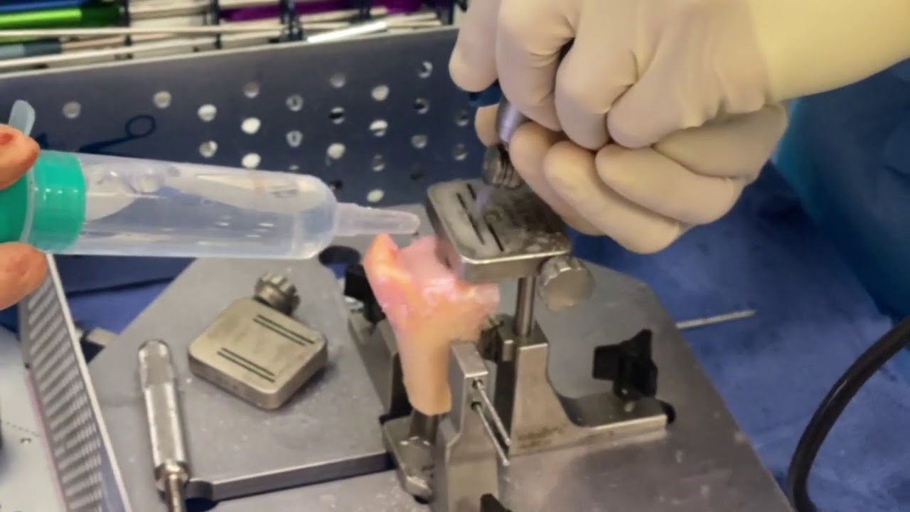

In this video, Dr. Grant Garcia demonstrates his open surgical technique for a Distal Tibial Allograft (DTA) shoulder reconstruction. This complex procedure is indicated for patients with severe, recurrent anterior shoulder instability who have failed previous soft tissue repairs and present with significant glenoid bone loss.
In this specific case, the patient had greater than 32% bone loss on the front of the shoulder socket (glenoid). Because the defect was so large, the patient was not a suitable candidate for a traditional Latarjet procedure (which uses a smaller bone block from the shoulder blade). Instead, a fresh osteochondral (bone-and-cartilage) distal tibial allograft from a donor was utilized to completely reconstruct the socket.
The key surgical stages demonstrated in the video include:
- Exposure and Tissue Dissection: An open deltopectoral approach is performed through a cosmetic incision along Langer's lines. The surgeon splits the subscapularis muscle horizontally (a "50-yard line split") and dissects it away from the underlying joint capsule. Because of significant scarring from a previous failed surgery, old suture anchors are carefully debrided and removed.
- Capsulotomy and Joint Retraction: The capsule is carefully incised and tagged with FiberWire sutures to protect the remaining native joint cartilage. A Fukuda retractor is placed to pull the humeral head out of the way, and specialized angled Gelpi and anterior Bankart retractors are deployed to achieve an expansive, intra-articular view of the damaged glenoid rim.
- Allograft Graft Preparation: Dr. Garcia uses a specialized guide system to shape the fresh donor bone graft to the precise measurements calculated on pre-operative 3D imaging. For this patient, he cuts a graft that is 7 mm thick, 10 mm deep, and 22 mm from superior to inferior, applying a 10-degree cutting offset to perfectly match the natural curvature of the native socket. Irrigation is performed continuously during the saw cuts to preserve the donor tissue.
- Marrow Clearing and PRP Preparation: The shaped allograft is thoroughly cleaned with pulse lavage to flush out donor marrow elements (which reduces immunogenic risks) and is then soaked in Platelet-Rich Plasma (PRP) to stimulate cellular integration and maximize biological bone healing.
- Graft Fixation and Compression: The graft is held against the prepared native glenoid wall using parallel guide pins. Dr. Garcia drills through the graft into the native bone and slides a specialized two-hole compression plate over the top. Fully threaded cannulated screws are driven through the plate, compressing the donor bone flush against the socket. An assistant uses a Freer elevator to ensure a completely smooth transition with zero step-off between the native cartilage and the allograft cartilage.
- Capsular Reconstruction: Before final tightening of the hardware, suture tapes are channeled underneath the compression plate. These heavy tapes are then woven through and used to securely tie down the scarred anterior capsule, reinforcing front-side stability.
A final intra-articular visual check confirms that the 32% bone defect has been entirely restored with an anatomically flush, fresh cartilage surface, effectively normalizing the shoulder socket footprint to eliminate the risk of future dislocations.







