

 Book An Appointment



Dr. Garcia demonstrates his innovate technique for meniscal root repair.
Dr. Garcia specializes in complex knee, shoulder and elbow sports surgeries. He has prepared a number of surgical videos below to help patients better understand their procedures. He is frequently updating his surgical video database so check back soon for further updates.
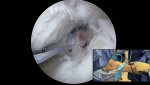

This video features Dr. Grant H. Garcia demonstrating his innovative arthroscopic surgical technique for repairing a medial meniscus root tear. Meniscus root repairs are critical for preserving joint mechanics and protecting the knee cartilage over time by restoring normal "hoop stresses" across the meniscus.
Here is a summary of the procedure shown in the video:
- Initial Visualization and Access: The procedure begins with a diagnostic arthroscopy to evaluate the knee and probe the detached medial meniscal root. To improve access within this notoriously tight joint space, the surgeon performs a trephination of the medial collateral ligament (MCL) using a spinal needle, allowing the joint to safely open up.
- Preparing the Footprint and Notch: A 4.5mm or 5.5mm shaver is used to perform a reverse notchplasty, taking out a small fragment of the posterior cruciate ligament (PCL) and widening the area. The surgeon debrides the scarred meniscal root and uses a curved curette to remove overlying articular cartilage at the native bony insertion footprint. A meniscal rasp is also used to prepare the tissue, exposing a raw bone and vascular bed necessary for the root to heal.
- Suture Placement (Cinch Stitches): A specialized cannula is placed into the knee to prevent suture tangling and "bridging" across tissues. Using a specialized curved tissue passer, the surgeon places a robust, structurally sound cinch stitch through the peripheral edge of the meniscus using a heavy-duty mini tape. A second cinch stitch is placed closer to the torn edge of the root using a different colored mini tape to keep the strands easy to identify.
- Tunnel Drilling: A targeted tibial guide system is locked onto the anatomic meniscal root footprint. A drill bit with a protective metal sleeve is advanced through the tibia. Once fat debris is observed (indicating proximity to the joint surface), the guide is removed, and the surgeon slowly drills through the final bone cortex under direct vision to prevent damaging the knee condyle or posterior structures.
- Graft Passing and Tensioning: A rigid wire suture-passer is inserted up through the tibial tunnel, grabbed through the medial portal, and used to pull all four limbs of the mini tape down through the bone tunnel. Holding the strands firmly to avoid bunching, the surgeon pulls the meniscal root flat against its anatomical footprint.
- Securing and Evaluating: With the knee positioned at 60 degrees of flexion, the surgeon completes two separate rounds of sequential tightening on the sutures. This ensures optimal tension and pulls the torn root tightly into the bone tunnel. The surgeon uses a probe to confirm that the construct is highly stable and that the anatomical gap is completely closed.
- Marrow Stimulation: Finally, the surgeon performs a microfracture procedure in the notch area. This introduces marrow elements and stem cells into the joint, which orthopedic data suggests improves meniscus-to-bone healing rates by roughly 25%.







