

 Book An Appointment



Dr. Garcia demonstrates his cartilage defect treatment using MACI.
Dr. Garcia specializes in complex knee, shoulder and elbow sports surgeries. He has prepared a number of surgical videos below to help patients better understand their procedures. He is frequently updating his surgical video database so check back soon for further updates.
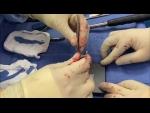

In this video, Dr. Grant H. Garcia demonstrates a MACI (Matrix-induced Autologous Chondrocyte Implantation) procedure to treat a trochlear cartilage defect in the knee. The patient previously underwent a diagnostic arthroscopy and biopsy to grow their own cartilage cells in a lab prior to this final implantation stage.
The surgical procedure involves the following steps:
- Defect Preparation: A medial arthrotomy is performed to open the knee and expose the moderate-sized cartilage defect. Dr. Garcia uses an angled curette to carefully clean out the damaged cartilage down to the calcified cartilage layer, taking strict care not to damage the subchondral bone.
- Hemostasis (Bleeding Control): The procedure is done off-tourniquet. Dr. Garcia uses epinephrine-soaked patties and a small amount of fibrin glue massaged into the capillaries to ensure there is zero bleeding or hematoma formation, which could ruin the graft.
- Graft Cutting and Orientation: A custom cutting guide is used to precisely size the MACI implant membrane to match the patient's defect. The surgeon identifies the "cell side" of the graft and handles it delicately with toothless forceps.
- Implantation and Fixation: A layer of fibrin glue is applied to the base of the cleaned bone defect. The MACI membrane is flipped so that the cell side faces down toward the bone defect (leaving the shiny side up). It is positioned into the lesion, and more fibrin glue is applied around the neatly rounded edges.
- Final Testing: The surgeon uses digital pressure (pressing down with a finger) for roughly three minutes to secure the fixation. Finally, the knee is cycled through a range of motion to ensure the graft stays firmly in place without any catching, shifting, or delamination.







