

 Book An Appointment



Dr. Garcia demonstrates his arthroscopic technique for knee osteochondritis dissecans (OCD) repair.
Dr. Garcia specializes in complex knee, shoulder and elbow sports surgeries. He has prepared a number of surgical videos below to help patients better understand their procedures. He is frequently updating his surgical video database so check back soon for further updates.
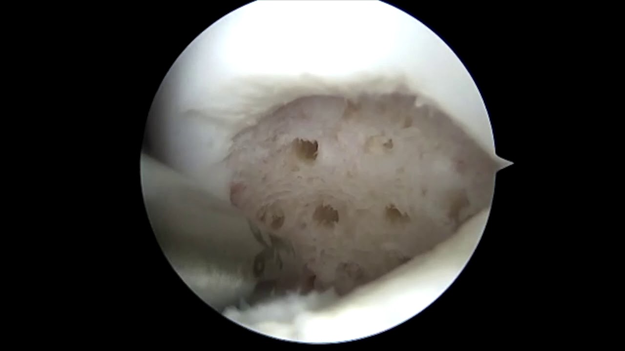
This surgical technique outlines a highly precise, minimally invasive method for treating unstable osteochondritis dissecans (OCD) lesions in the knee. The goal of this arthroscopic procedure is joint preservation—specifically salvaging and re-fixing the patient’s own detached or loose cartilage-and-bone fragment back onto the femoral condyle rather than removing it.
Here is a summary of the technical workflow used for an arthroscopic knee OCD repair:
- Diagnostic Evaluation and Fragment Assessment: The procedure begins with a standard knee arthroscopy through specialized portals. The surgeon carefully introduces a probe to assess the stability, size, and tissue quality of the OCD lesion on the femoral condyle. If the fragment is unstable but structurally intact, the decision is made to perform a primary internal fixation.
- Bed Preparation and Microfracture/Drilling: The loose cartilage and bone fragment is gently elevated or hinged open to expose the underlying damaged bone bed (the "crater"). The surgeon debrides any dead or sclerotic (hardened) bone and scar tissue from both the underside of the fragment and the base socket using a small shaver or curette. To bring fresh blood flow and healing marrow elements to the injury site, multiple small holes are drilled or a microfracture technique is performed within the exposed bone bed.
- Anatomic Reduction: The loose OCD fragment is carefully repositioned and aligned back down into its native anatomical bed. The surgeon ensures the borders match up flawlessly to recreate a perfectly smooth, congruent cartilage profile along the weight-bearing surface of the knee joint.
- Compression and Internal Fixation: While holding the fragment rigidly in place, guide pins are drilled across the cartilage piece and deep into the underlying bone. The fragment is then secured using specialized compression devices—frequently bioabsorbable smart-screws, pins, or low-profile headless metal compression screws. These fasteners are inserted flush or slightly countersunk below the joint cartilage surface to protect the opposite side of the knee from friction.
- Stability Testing: The surgeon uses an arthroscopic probe to aggressively stress the edges of the repaired fragment, verifying that the mechanical fixation is entirely rigid and will not displace during early knee movement. The knee joint is passed through a full range of motion to ensure there is no catching or micro-motion before the surgical instruments are withdrawn and the minimal incisions are closed.







