

 Book An Appointment



Dr. Garcia demonstrates his arthroscopic surgical technique for anterior labrum “Bankart”
Dr. Garcia specializes in complex knee, shoulder and elbow sports surgeries. He has prepared a number of surgical videos below to help patients better understand their procedures. He is frequently updating his surgical video database so check back soon for further updates.
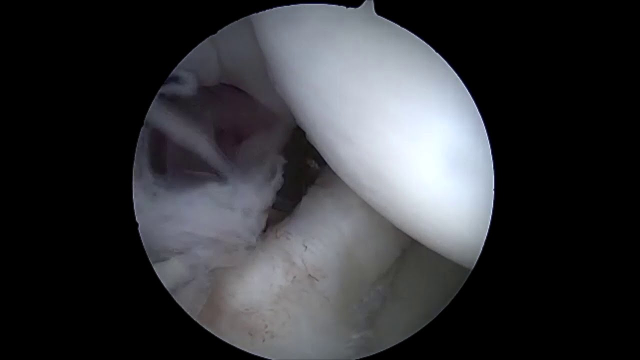
In this surgical video, Dr. Grant Garcia demonstrates his advanced arthroscopic technique for an anterior labrum "Bankart" repair. The patient has a significant anterior labral tear, accompanied by a small cartilage defect and a GLAD (Glenoid Labral Articular Disruption) lesion caused by multiple shoulder dislocations.
The demonstration covers the following key surgical stages:
- Diagnostic Evaluation and Portal Placement: Following an initial diagnostic arthroscopy to evaluate the extent of the tissue damage, Dr. Garcia establishes working access. He maps out three distinct portals: a posterior portal for grasping, an anterior-superior portal for main instrument manipulation, and a specialized percutaneous trans-subscapularis (trans-subscap) portal. A small cannula is used in the trans-subscap portal to minimize muscle trauma.
- Tissue Mobilization and Bed Preparation: Using a Bankart elevator and a rasp, the surgeon carefully mobilizes the detached labrum and aggressively rasps the anterior glenoid bone. Preparing a bleeding bone bed is a critical step to optimize the biological healing potential of the reattached soft tissue.
- Six O'Clock Anchor Fixation: Dr. Garcia passes a specialized suture tape anchor (Arthrex Suture Tack) low down at the 6 o'clock position on the glenoid rim. Data supports utilizing a trans-subscap portal to safely achieve this low placement, which is linked to lower recurrence rates. He weaves the robust suture tape through the inferior glenohumeral ligament in a horizontal mattress fashion, tying it with alternating half-hitches to reconstruct a flat, thick bumper that stands clear of the cartilage.
- Four O'Clock and Three-Thirty Fixation: Moving sequentially upward, a second Suture Tack is deployed at the 4 o'clock position, and a third is placed around the 3:30 position. Both are passed through the tissue and tied down using a horizontal mattress configuration. This specific pattern avoids placing large prominent knots directly on the articular cartilage while compressing the labrum flat against the prepared bone bed.
- Construct Evaluation: The completed labral-capsular repair is checked thoroughly from the anterior-superior portal view. Dr. Garcia uses an arthroscopic probe to physically manipulate the newly constructed tissue bumper, confirming that it is tightly adhered to the bone and provides structural resistance against translation.
Dr. Garcia emphasizes that this procedure is performed with the patient in the lateral decubitus (side-lying) position, rather than a standard beach-chair position, as clinical evidence points to a lower post-operative redislocation rate when utilizing traction in this orientation. A final manual examination under anesthesia confirms the shoulder is entirely stable with no residual anterior sliding or subluxation.







