

 Book An Appointment



Dr. Garcia demonstrates his arthroscopic remplissage technique
Dr. Garcia specializes in complex knee, shoulder and elbow sports surgeries. He has prepared a number of surgical videos below to help patients better understand their procedures. He is frequently updating his surgical video database so check back soon for further updates.
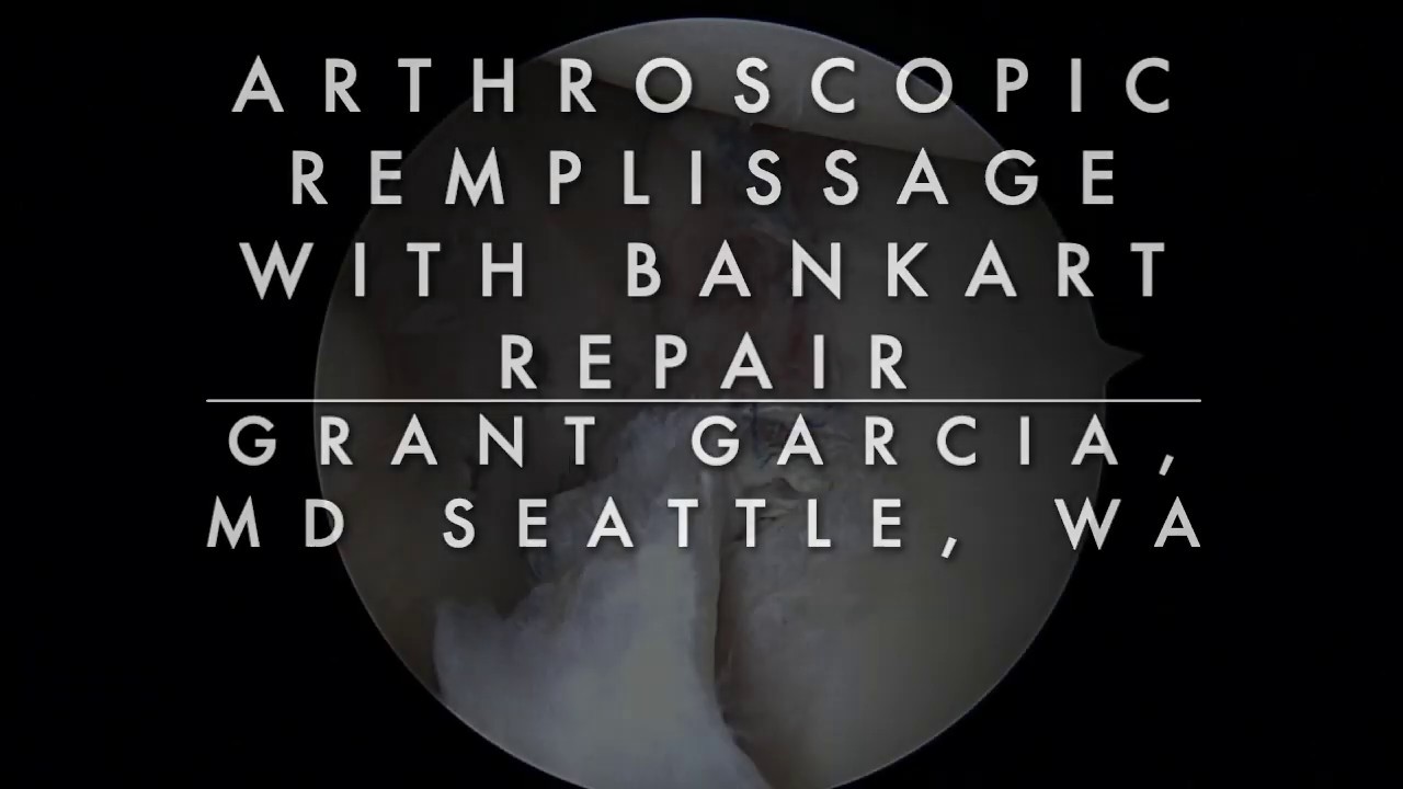

In this video demonstration, Dr. Grant Garcia highlights his surgical technique for an arthroscopic Remplissage combined with a revision Bankart repair. The patient previously underwent a failed Bankart repair by an outside surgeon, resulting in recurrent instability and a large, deep "off-track" Hill-Sachs lesion (a bone dent on the back of the humeral head) calculated via 3D CT imaging.
The Remplissage technique is designed to "fill" this bone defect by anchoring the adjacent infraspinatus tendon and shoulder capsule directly into the dent, preventing it from catching on the socket rim.
The demonstration outlines the following key stages of the procedure:
- Hill-Sachs Defect Preparation: Viewing from a posterior approach, Dr. Garcia uses a curette and a surgical shaver to clear away tissue from the deep Hill-Sachs bone dent. He rasps the base down to a fresh, bleeding bony bed to promote strong biological healing when the tendon is compressed into the socket.
- Anchor Placement: He deploys two 2.6 mm knotless FiberTack anchors loaded with heavy suture tape directly into the base of the bone defect. The anchors are spaced roughly 1.5 centimeters apart along a superior-to-inferior line.
- Percutaneous Suture Passing: Working percutaneously from outside the shoulder, Dr. Garcia inserts a 17-gauge spinal needle at a 45-degree angle to pierce through the infraspinatus tendon. He passes a nitinol (NiTi) wire loop through the needle to grab and shuttle the suture strands from the lower anchor through the deep surface of the tendon. He repeats this process 1 to 1.5 centimeters higher up to retrieve the sutures from the top anchor.
- Subacromial Space Clearing: Protective "fiber sticks" are temporarily slid over the external suture strands. This safeguards the repair lines while the surgeon shifts into the subacromial space to clean out bursa and scar tissue, ensuring a clear path for final knotless tightening.
- Knotless Tensioning and Tissue Engagement: The surgeon confirms that the sutures are anchored firmly in the robust tendon tissue rather than muscle belly. He then threads the heavy suture tapes into the self-locking loops of the knotless anchors.
- Sequential Cinching: While the video focuses on the final cinching, Dr. Garcia explains the structural order of operations: the Remplissage anchors are placed first, the revision anterior Bankart labral repair is completed to address front-side stability, and then both Remplissage anchors are sequentially cinched down tightly from the back.
This synchronized tensioning draws the infraspinatus tendon flat into the Hill-Sachs cavity, establishing a secure "suction fit" that fills the structural bone defect and eliminates the risk of future engaging shoulder dislocations.







