

 Book An Appointment



Dr. Garcia demonstrates his advanced technique for arthroscopic glenoid fracture fixation ...
Dr. Garcia specializes in complex knee, shoulder and elbow sports surgeries. He has prepared a number of surgical videos below to help patients better understand their procedures. He is frequently updating his surgical video database so check back soon for further updates.
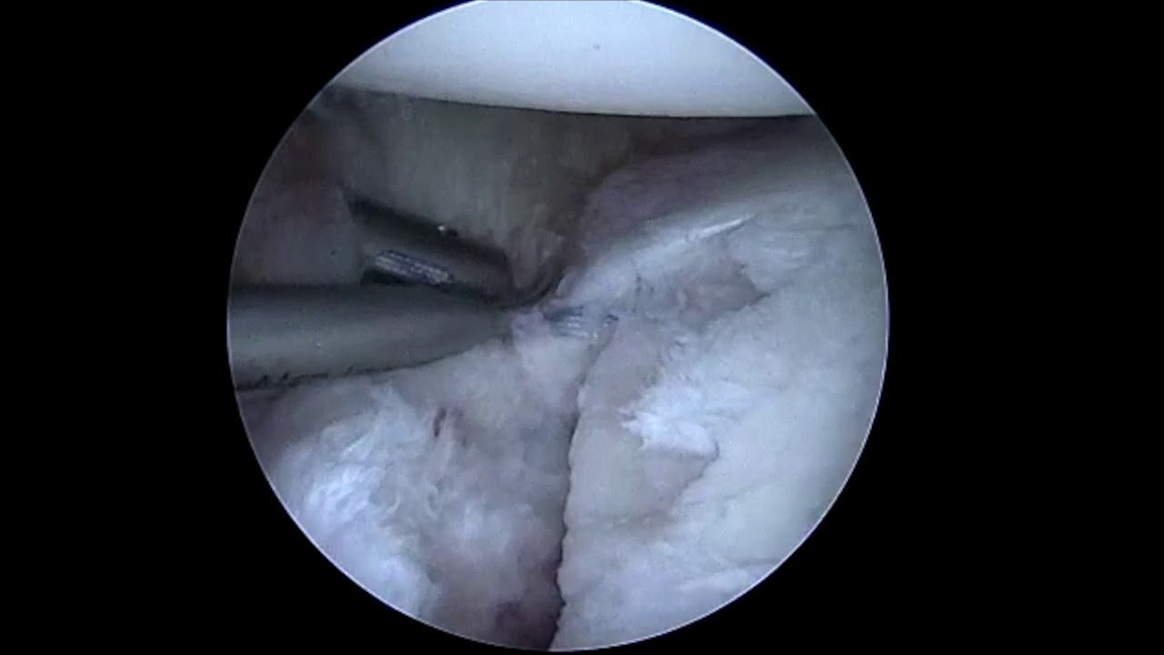

In this orthopedic demonstration video, Dr. Grant Garcia shares his advanced, minimally invasive arthroscopic technique for fixing a glenoid fracture. This specialized approach can be utilized to treat acute bone fractures of the shoulder socket or significant bony Bankart injuries (where a piece of the socket's rim breaks off during a shoulder dislocation). In this specific case, the patient has a large fracture covering greater than 25% of the glenoid surface with a 3 mm structural displacement or step-off.
The demonstration breaks down into the following key surgical phases:
- Fracture Site Debridement: Using an arthroscopic shaver, Dr. Garcia carefully cleans out the fracture gap, clear away loose debris, and preps the bone to optimize its healing potential. Throughout this, he is meticulous about saving all healthy articular cartilage and the surrounding labrum.
- Percutaneous Portal Creation: To reach the area safely, he creates a specialized trans-subscapularis portal using a spinal needle guide and a small 5 mm cannula. This minimizes muscle trauma to the critical subscapularis tendon on the front of the shoulder.
- Medial Anchor Placement: To start building the fixation construct, a medial suture anchor is placed into the main bone socket, resting just inside (medial to) the detached fracture fragment.
- Horizontal Mattress Suture Passing: Using a curved Crescent Lasso instrument, Dr. Garcia individually passes heavy suture tapes under and around the fracture fragment. By looping these in a horizontal mattress pattern, he creates an underlying suture sling. This step is technically demanding but vital—it pulls the floating bone fragment up into place and prevents it from flipping inward (inverting) during final tightening.
- Inferior and Superior Labral Repair: Turning his attention to soft tissue stability, the surgeon places additional anchors (Suture Tacks) at the lower and upper margins of the labrum. He captures a sufficient amount of the inferior glenohumeral ligament and ties them down with alternating half-hitches. This re-establishes a robust, elevated soft tissue bumper around the newly aligned bone fragment.
- Compression and Final Fixation: To fully cinch the broken bone pieces back together, the remaining suture tails from the original medial anchor are brought across the fracture line. They are fed into an anterior PushLock anchor and impacted into the bone. This final step applies deep, structural compression across the fracture bed.
At the close of the procedure, a probe test confirms that the fragment is tightly realigned, flush with the rest of the joint surface, and displays absolutely zero movement or shifting under manual pressure.







