

 Book An Appointment



Dr. Garcia demonstrates first live MACI Arthro video in the US
Dr. Garcia specializes in complex knee, shoulder and elbow sports surgeries. He has prepared a number of surgical videos below to help patients better understand their procedures. He is frequently updating his surgical video database so check back soon for further updates.
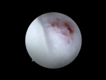

This video features Dr. Garcia demonstrating a MACI (Matrix-induced Autologous Chondrocyte Implantation) Arthro procedure. It is highlighted as a primary demonstration of an entirely arthroscopic MACI procedure in the United States using a dedicated instrumentation set to treat a trochlear cartilage lesion.
The surgical procedure involves the following steps:
- Portal Creation and Access: The surgeon identifies the proper trajectory using a spinal needle. Radiofrequency (RF) energy is utilized to minimize bleeding across the working areas. Medial and lateral portals are established, followed by a transpatellar portal (less than 2 cm long) created while the knee is in slight flexion to cleanly split the tendon. A large 15 mm cannula is inserted through this central portal.
- Defect Preparation: A small cutting guide is inserted through the cannula, wiggled through the cartilage layer, and gently malleted to outline the boundaries of the lesion. Working meticulously through all three portals, the surgeon uses various curettes, rongeurs, and a shaver to clear away the damaged tissue down to the calcified cartilage layer, taking care not to compromise healthy bone or leave jagged cartilage edges.
- Hemostasis (Drying the Joint): For a MACI implant to adhere, the target site must be completely dry. The surgeon switches to a "dry scope" technique, completely deflates the tourniquet, and handles bleeding using epinephrine-soaked patties, a Fraser suction tip, and topical gel thrombin.
- Graft Cutting and Loading: On the back table, the surgeon utilizes a disposable, sharp matching cutter to punch out a custom-sized graft from the patient's cultured MACI membrane. The graft is carefully loaded—cell side up—onto a specialized V-shuttle tool, which secures the membrane with an advanced suction seal.
- Implantation and Sealing: A thin foundation layer of fibrin sealant (Tisseel) is applied to the bone using a spinal needle applicator. The V-shuttle is passed into the knee, the graft is dropped over the defect, and the tool's prongs are released. The surgeon uses a specialized plastic-tipped probe to smooth out the graft, eliminate air pockets, and compress the edges.
- Final Verification: After applying a top layer of fibrin sealant and letting it set for three minutes, the surgeon takes the knee through a manual range of motion. The implant demonstrates excellent stability without shifting or catching, paving the way for the patient to begin immediate postoperative rehabilitation.







