

 Book An Appointment



Dr. Garcia demonstrates an ACL repair around a tibial nail
Dr. Garcia specializes in complex knee, shoulder and elbow sports surgeries. He has prepared a number of surgical videos below to help patients better understand their procedures. He is frequently updating his surgical video database so check back soon for further updates.
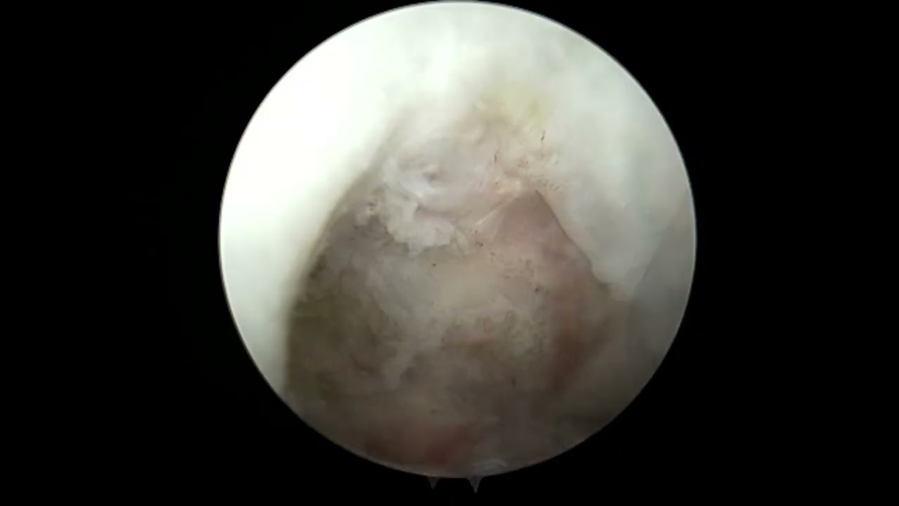
In this unique surgical video, Dr. Grant H. Garcia presents a highly creative workaround: performing a primary ACL repair enhanced with an internal brace on a patient who has an indwelling intramedullary tibial nail. The nail had been placed at an outside facility 11 weeks prior to manage a tibial shaft fracture and cannot be removed until the bone fully heals.
Because standard ACL reconstruction tunnels would collide with the metal nail, Dr. Garcia opts for a primary repair with a specialized drilling trajectory:
The procedure follows these precise phases:
- Notch Preparation & Stitching: The procedure begins by microfracturing the femoral notch to stimulate bone marrow stem cells and promote vascular ingrowth. Dr. Garcia passes 2.5 mm FiberRings through the torn ACL tissue. The 25 mm loop size is specifically chosen to accommodate the robust size of the patient's native tissue and prevent any suture slippage.
- Navigating Around the Nail: To establish the tibial tunnel for the internal brace, Dr. Garcia uses a bullseye guide. The drill must veer slightly more medial than normal to bypass the hardware. On the first pass, the drill actually skims the tibial nail, requiring an intraoperative adjustment to angle further medially to achieve clear passage.
- Femoral Drilling & Button Inserter: A spade-tip drill is advanced through the femoral cortex, cycling back and forth to ensure the channel is clean. A passing stitch is pulled out through the lateral portal. A specialized TightRope construct (similar to a bone-patellar tendon-bone TightRope) is pre-loaded with the internal brace and the FiberRing sutures.
- Seating the Graft & Internal Brace: The femoral button is pulled up and flipped onto the outer femoral cortex. Dr. Garcia holds manual back-tension on the internal brace strands during this process to ensure they do not accidentally migrate or catch on the iliotibial (IT) band.
- Tensioning & Final Stability: Once the button is flipped, the white TightRope sutures are incrementally tensioned to draw the torn native ACL tightly up against the bleeding bone of the femoral notch. The internal brace is then brought down into the adjusted tibial tunnel.
The final assessment shows excellent native tissue vascularity and superb structural tension. On physical examination, the knee's anterior stability is completely restored, and the Lachman test successfully returns to normal.







