

 Book An Appointment



Dr. Garcia demonstrates a cartilage transplant surgery.
Dr. Garcia specializes in complex knee, shoulder and elbow sports surgeries. He has prepared a number of surgical videos below to help patients better understand their procedures. He is frequently updating his surgical video database so check back soon for further updates.
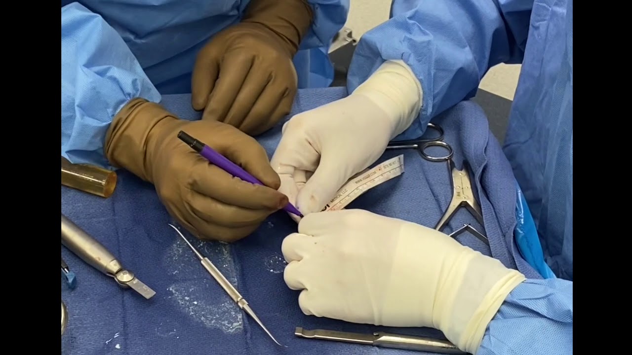
This video features Dr. Grant H. Garcia demonstrating a highly specialized cartilage transplant surgery known as an Osteochondral Allograft (OCA) transplantation of the trochlea. Trochlear cartilage transplants are uniquely complex because of the groove's intricate, three-dimensional curvature, which requires matching the recipient's anatomy across multiple planes to prevent the patella (kneecap) from catching or tracking incorrectly.
Here is a summary of the technical workflow used for this cartilage transplantation, presented without timestamps:
- Sizing and Recipient Bed Preparation: The surgeon uses calibrated sizing rings directly over the cartilage defect to determine the exact diameter required for a secure press-fit wedge. In this case, a 22.5mm target socket is established. A guide pin is drilled into the center of the defect, and a specialized reamer is advanced sequentially. Because the trochlea has a sloped surface, all four quadrants of the socket generally display distinct depths. The surgeon carefully gauges the depth to ensure the shallowest edge reaches at least 5 millimeters. The guide wire remains in place to preserve the perfect orientation angle.
- Harvesting the Donor Graft: The donor distal femur allograft is locked into a rigid workstation. The surgeon aligns the donor anatomy to match the orientation of the patient's knee. Using a coring reamer under copious cold irrigation, a cylindrical bone-and-cartilage plug is drilled to a depth of roughly 20 millimeters. A surgical saw is then used to cut and free the plug from the donor bone. A blunt elevator is carefully utilized to handle the graft, ensuring nothing touches or damages the delicate cartilage surface.
- Precision Matching and Sculpting: To replicate the recipient's sloped bone bed, the surgeon measures the exact depth of the patient's socket at the 12, 3, 6, and 9 o'clock positions. These four individual measurements are mapped directly onto the donor plug, and the markings are connected. The plug is securely held in a cutting jig while a saw removes the excess bottom bone, tailoring it to fit perfectly flat. A "waffle cut" grid pattern is scored into the bottom bone face to provide a slight margin of error for compression. The bottom edge of the plug is lightly chamfered to assist with initial entry into the tight recipient socket.
- Preparing the Biological Environment: Small 2.0mm subchondral holes are drilled into the floor of the recipient bed to encourage marrow element migration. Both the recipient bone socket and the donor plug undergo thorough pulse lavage to wash away free marrow elements and target red blood cell debris. To maximize healing potential, platelet-rich plasma (PRP) is harvested from the patient and mixed with a demineralized bone matrix (DBM) putty, which is packed smoothly into the base holes. The remaining liquid PRP is soaked directly into the porous bone base of the donor graft.







