

 Book An Appointment



Complex vertical and horizontal meniscus repair
Dr. Garcia specializes in complex knee, shoulder and elbow sports surgeries. He has prepared a number of surgical videos below to help patients better understand their procedures. He is frequently updating his surgical video database so check back soon for further updates.
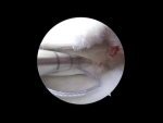
This video, titled "Complex Vertical and Horizontal Meniscus Repair," demonstrates an arthroscopic technique to repair a multifaceted meniscus injury in a young patient. The patient has a large vertical bucket-handle tear (where the inner part flips into the center of the joint like a bucket handle) combined with a deep horizontal component near the capsular attachment.
To achieve maximum compression and stability across both tear directions, the surgeon combines all-inside anchors, inside-out suturing, and specialized horizontal repair devices.
The primary surgical steps shown in the video include:
- Tear Reduction and Debridement: The surgeon first physically flips the displaced bucket-handle component back into its correct anatomical position (reducing the tear). A shaver is used to debride and clean the torn tissue edges to prepare them for healing. A passport cannula is placed to keep portals open and prevent sutures from catching on the surrounding soft tissues.
- Initial All-Inside Fixation: To anchor the tissue near the meniscus root and prevent it from immediately flipping back out of place, an all-inside anchor (Arthrex FiberStitch) is deployed. This device utilizes two robust suture limbs to provide excellent initial pull-out strength.
- Inside-Out Vertical Repair: Turning attention to the lengthy vertical component of the tear, the surgeon uses an inside-out technique. Suture tape needles are passed through the joint and out an accessory incision, placing multiple vertical mattress sutures. This pattern offers the highest possible biomechanical strength and compresses the main body of the meniscus securely back against its outer capsule.
- Managing the Horizontal Component: With the vertical tear secured, the surgeon addresses the secondary horizontal cleavage component running through the posterior horn. A specialized suture passer is angled underneath both the upper and lower leaflets of the tear. This allows the surgeon to pass sutures directly through the layers without striking the nearby femoral joint cartilage.
- Knot Tying and Compression: The horizontal sutures are tied down sequentially using a series of alternating half-hitches. The surgeon ensures the knots are buried cleanly underneath the meniscus body to protect the joint surfaces. Additional inferior sutures are added to lock down the bottom flap, ensuring zero residual mobility when probed.
- Biologic Augmentation (Notch Microfracture): After verifying a rock-solid, uniform repair, the surgeon performs microfracturing in the femoral notch. This step mimics the bone-bleeding response of an ACL tear, releasing a rush of fresh bone marrow and stem cells into the joint fluid to dramatically boost the healing potential of the repaired meniscus.







