

 Book An Appointment



ACL repair, MCL repair, Lateral meniscal root repair
Dr. Garcia specializes in complex knee, shoulder and elbow sports surgeries. He has prepared a number of surgical videos below to help patients better understand their procedures. He is frequently updating his surgical video database so check back soon for further updates.
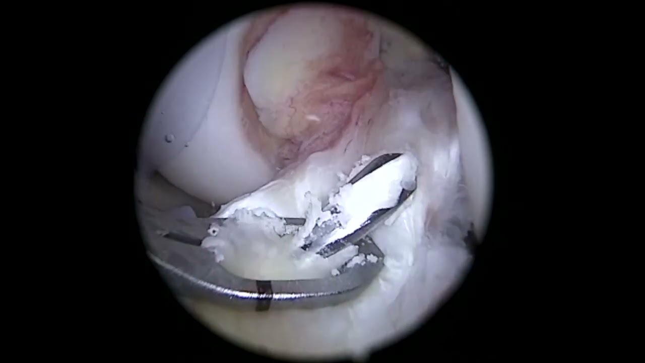
This video, titled "ACL repair, MCL repair, Lateral meniscal root repair," details a highly complex multi-ligament knee reconstruction and repair. The patient has structural hyperlaxity (naturally loose joints) and suffered a severe multi-injury trauma consisting of a proximal ACL tear, a Grade 3 Medial Collateral Ligament (MCL) tear with over 6mm of abnormal joint gapping, and a lateral meniscal root tear.
To address all three issues effectively without removing any of the patient's native, highly vascular tissues, the surgeon combines specialized all-inside, transtibial, and internal bracing techniques.
The core phases of this complex multi-step procedure include:
- Preparation and Notch Microfracture: The surgeon performs a minor notchplasty to prepare the area, clearing away any scarred tissue surrounding the torn ACL stump. The femoral notch is microfractured to introduce a rush of fresh blood and stem cells to boost the healing potential of the repair. A 4.5mm pin is then drilled completely through the femoral wall.
- Lateral Meniscal Root Repair (Transtibial Tunnel): The surgeon addresses the lateral meniscal root and radial tear flap. A ball rasp is used to irritate the bone footprint to encourage blood flow. Two separate sutures are passed through the meniscus: a simple link suture and a horizontal mattress suture. A dedicated transtibial bone tunnel is drilled beneath the root site, and the suture limbs are pulled straight down through this tunnel to anchor the meniscus securely back to the bone.
- Secondary All-Inside Meniscal Fixation: To prevent the newly anchored meniscus from extruding out of the side of the joint, the surgeon reinforces the repair with an all-inside anchor (FiberStitch). This provides a secondary loop of support that bridges from the root to the outer periphery, creating a highly robust fixation network.
- ACL Repair with Internal Brace: Turning back to the ACL, the surgeon loops a 35mm FiberRing suture system through the patient’s thick, vascular native tissue stump. The sutures are connected to an adjustable cortical button system and threaded up into the femoral tunnel. The button is flipped flat on the outer bone wall (verified via X-ray), and a high-strength suture tape (internal brace) is added for structural reinforcement.
- Tensioning and Stability Check: The surgeon tightens the adjustable loops to pull the native ACL stump firmly back against its original anatomical footprint. A physical test (Lachman test) confirms that normal joint tightness has completely returned.
- MCL Repair Validation: Though the physical cutting and pinning of the MCL repair is not explicitly shown on camera, the surgeon anchors the MCL using a secondary internal brace construct. A final stress test confirms that the severe 6mm inner knee gapping has been successfully reduced down to a stable 3mm, completing the combined multi-ligament reconstruction.







