

 Book An Appointment



ACL Patella Tendon Reconstruction with Tightrope and Internal Brace.
Dr. Garcia specializes in complex knee, shoulder and elbow sports surgeries. He has prepared a number of surgical videos below to help patients better understand their procedures. He is frequently updating his surgical video database so check back soon for further updates.
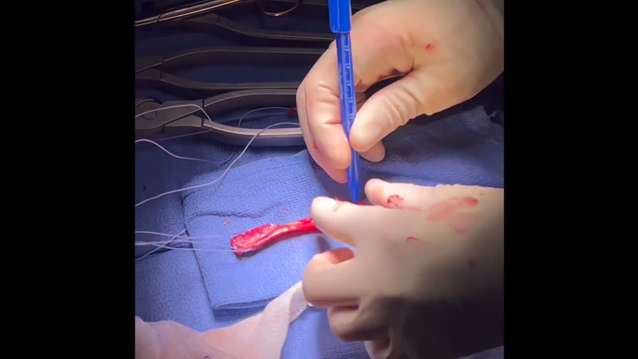

This video, titled "ACL Patella Tendon Reconstruction with Tightrope and Internal Brace," demonstrates an advanced arthroscopic ACL reconstruction technique typically used for younger, active patients. It utilizes a patellar tendon autograft (the patient's own tissue with bone plugs on either end) but replaces traditional rigid screw fixation on the femur with an adjustable TightRope button system and an internal brace (high-strength suture tape).
This combination gives the surgeon adjustable tensioning capabilities and adds structural reinforcement that is difficult to achieve with standard screw-only setups.
The key surgical steps demonstrated in the video include:
- Femoral Tunnel Preparation: Using a 7mm over-the-top guide, the surgeon reams a socket into the femur to a depth of about 25mm. A 4.5mm drill then passes all the way through the outer bone cortex to establish the path for the fixation button. The tunnel is completely cleaned to ensure the bone plug can slide in smoothly.
- Tibial Tunnel Drilling: An elbow guide is used to precisely align the tibial (shinbone) tunnel, which is then opened up with a straight reamer.
- Graft Preparation & TightRope Attachment: On the back table, the patellar tendon bone plugs are measured, trimmed, and crimped to guarantee smooth passage. Suture loops are woven into the tibial side bone plug. For the femoral side, the new BT (Bone Tendon) TightRope system is attached to the bone block, ensuring the high-strength suture tape sits securely at least 5mm from the edge of the bone plug. The tensioning strands are pulled to ensure they are completely even.
- Graft Passage & Button Flipping: A passing suture pulls the graft assembly up through the tibial tunnel and into the femoral socket. The surgeon watches for the femoral cortical button to clear the bone wall and flip flat against the outside of the thigh bone, which is verified using intraoperative X-ray.
- Seating the Bone Plug: Because it is a rigid bone plug rather than soft tissue, it doesn't just pull up effortlessly. The surgeon uses a straight snap instrument to carefully guide the plug into the femoral socket, ensuring the smooth cortical side faces backward (posteriorly) until it is fully seated.
- Tibial Fixation and Final Check: With the knee held completely straight, an interference screw is driven into the tibial tunnel to lock the bottom bone plug in place. Simultaneously, the internal brace suture tape is anchored to the bone for secondary reinforcement.
The surgeon finishes with a physical stability test (Lachman test), demonstrating that the knee has minimal laxity and has successfully returned to normal structural stability.







