

 Book An Appointment



AutoCart Cartilage Replacement
AutoCart Cartilage Restoration Surgery in Seattle
Advanced, Single-Stage Cartilage Repair Using Your Own Cartilage
Introduction: A New Approach to Cartilage Restoration
Articular cartilage injuries of the knee can cause persistent pain, swelling, stiffness, and limitations in physical activity. These injuries commonly occur in athletes and active individuals following trauma, repetitive loading, or sports-related injuries. Because articular cartilage has a limited ability to heal naturally, significant cartilage defects can remain symptomatic despite physical therapy and other nonsurgical treatments.
Advances in orthopedic sports medicine have introduced innovative biological techniques designed to restore damaged cartilage while preserving the natural knee joint.
AutoCart is a single-stage cartilage restoration procedure that utilizes a patient’s own cartilage cells to treat localized cartilage defects. The technique involves harvesting healthy cartilage, processing it into small fragments, and implanting those fragments into the damaged area to support cartilage repair.
Unlike procedures requiring extensive cartilage transplantation or multiple operations, AutoCart allows cartilage harvesting and implantation during a single surgical procedure.
For appropriately selected patients, this approach offers a promising option for treating focal cartilage injuries while preserving the surrounding healthy joint.
What Is AutoCart?
AutoCart is an advanced cartilage restoration technique based on minced autologous cartilage implantation.
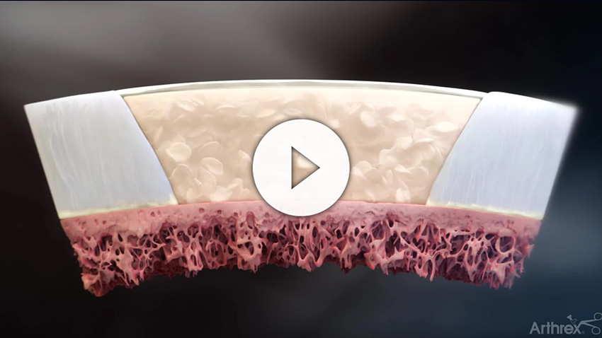
The procedure uses healthy articular cartilage obtained from the patient’s own knee. The cartilage is mechanically fragmented into small pieces, preserving viable cartilage cells known as chondrocytes.
These cells play an essential role in producing the extracellular matrix responsible for cartilage structure, durability, and mechanical function.
By creating numerous small cartilage fragments, surgeons increase the exposed cartilage surface area, potentially facilitating chondrocyte migration and biological repair.
The fragments are placed into the cartilage defect and stabilized using a biologically compatible fixation method, commonly fibrin sealant.
The objective is to encourage the formation of cartilage-like repair tissue that integrates with the surrounding joint surface.
AutoCart is particularly appealing because it combines biological cartilage restoration with a single-stage surgical approach.
However, although early clinical research is encouraging, long-term outcomes continue to be investigated.
Understanding Knee Cartilage Injuries
Articular cartilage is the smooth, specialized tissue covering the ends of bones within the knee joint. It allows nearly frictionless movement and helps distribute forces during walking, running, jumping, and athletic activities.
Unlike bone or muscle, cartilage has no direct blood supply. This limits its ability to regenerate following significant injury.
Common causes of cartilage damage include:
- Sports-related twisting or impact injuries
- Patellar dislocations
- Repetitive joint loading
- Previous ligament or meniscus injuries
- Osteochondral fractures
- Traumatic cartilage defects
Patients may experience activity-related pain, recurrent swelling, catching, stiffness, or difficulty returning to sports.
Cartilage injuries vary considerably in size, depth, and location. Some involve only the cartilage surface, while others extend into the underlying bone.
A comprehensive evaluation typically includes a physical examination, weightbearing radiographs, and MRI.
Treatment decisions depend on the characteristics of the defect, overall joint health, associated injuries, and patient activity goals.
Who Is a Candidate for AutoCart?
AutoCart may be considered for patients with symptomatic, localized cartilage defects who have not improved sufficiently with appropriate nonsurgical treatment.
Potential candidates include active adults with focal cartilage injuries involving the femoral condyles, trochlea, or patella.
The ideal candidate generally has a well-defined cartilage defect surrounded by healthy cartilage and adequate underlying bone support.
Important factors include:
- Defect characteristics: Size, location, depth, and condition of the underlying bone.
- Knee stability: Ligament injuries may need to be addressed to protect the cartilage repair.
- Meniscus integrity: Meniscus deficiency can increase forces across the cartilage defect.
- Lower-extremity alignment: Significant varus or valgus alignment may require correction.
- Overall joint health: Patients with advanced, diffuse osteoarthritis are generally not ideal candidates.
- Rehabilitation commitment: Successful recovery requires adherence to postoperative restrictions and physical therapy.
AutoCart is not appropriate for every cartilage injury. Selecting the correct procedure requires evaluating the entire knee rather than focusing exclusively on the cartilage defect.
Benefits of AutoCart Cartilage Restoration
AutoCart offers several potential advantages for appropriately selected patients.
1. Single-Stage Surgical Procedure
One of the primary benefits of AutoCart is that cartilage harvesting, preparation, and implantation occur during one operation.
This eliminates the need for a separate planned cartilage-harvesting procedure and laboratory cell cultivation.
A single-stage approach may simplify treatment and reduce the time between diagnosis and definitive cartilage restoration.
2. Uses the Patient’s Own Cartilage
AutoCart utilizes autologous cartilage containing living chondrocytes.
Because the cartilage comes from the patient, donor tissue is not required.
The technique preserves viable cartilage cells and portions of their surrounding extracellular matrix, potentially supporting biological repair.
3. Supports Biological Cartilage Repair
Laboratory research has demonstrated that viable chondrocytes can migrate from minced cartilage fragments and participate in extracellular matrix production.
The goal is to develop repair tissue capable of improving the damaged joint surface.
However, the resulting tissue may differ from normal native cartilage, and complete restoration of original cartilage structure cannot be guaranteed.
4. Minimally Invasive Surgical Options
Depending on the defect’s location and size, AutoCart may be performed arthroscopically or through a limited-open approach.
Smaller surgical exposures may reduce soft-tissue disruption while allowing accurate preparation and implantation.
The surgical approach is individualized to ensure adequate visualization and stable graft fixation.
5. Preserves the Natural Knee Joint
AutoCart is designed to treat focal cartilage damage while preserving native joint anatomy.
For selected patients, successful cartilage restoration may reduce symptoms, improve function, and support continued participation in recreational activities.
However, there is currently insufficient evidence to conclude that AutoCart prevents osteoarthritis or reliably delays knee replacement.
Scientific Evidence Behind AutoCart
The biological foundation of minced cartilage implantation has been investigated for more than two decades.
Early experimental research demonstrated that cartilage fragments contain viable chondrocytes capable of migrating into surrounding tissue and producing cartilage extracellular matrix.
Lu and colleagues demonstrated cartilage repair potential using minced cartilage tissue in a preclinical model, supporting the concept of fragment-based cartilage restoration.[1]
Subsequent research has examined cartilage fragment viability, cellular outgrowth, and the use of biological scaffolds to support cartilage regeneration.
Clinical investigations of autologous minced cartilage implantation have reported improvements in pain and patient-reported knee function in selected patients.
Nevertheless, the available clinical literature remains limited by relatively small study populations, variable surgical techniques, and incomplete long-term follow-up.
Current evidence supports AutoCart as a biologically promising cartilage restoration technique, but additional high-quality comparative studies are needed to establish its long-term effectiveness and durability.
How Is AutoCart Surgery Performed?
AutoCart is generally performed as an outpatient procedure. The surgical technique is tailored to the location and characteristics of the cartilage defect.
Step 1: Arthroscopic Evaluation
The procedure begins with a diagnostic knee arthroscopy.
A small camera is inserted into the joint to evaluate cartilage surfaces, ligaments, menisci, and surrounding structures.
The surgeon confirms the dimensions and depth of the cartilage defect and identifies any associated abnormalities.
Step 2: Cartilage Defect Preparation
Damaged and unstable cartilage is carefully removed.
The goal is to establish healthy, stable cartilage margins while preserving appropriate underlying bone.
Meticulous preparation is important to support graft placement and integration.
Step 3: Cartilage Harvesting
Healthy cartilage is harvested from an appropriate noncritical region of the knee or obtained from suitable cartilage tissue available during defect preparation.
The amount of cartilage collected depends on the size of the defect.
Care is taken to minimize unnecessary damage to healthy joint surfaces.
Step 4: Cartilage Fragment Preparation
The harvested cartilage is mechanically processed into small fragments.
Fragmentation increases the exposed surface area of the cartilage and may facilitate migration of viable chondrocytes.
The fragments are prepared for implantation according to the surgical technique.
Step 5: Cartilage Implantation
The prepared cartilage fragments are distributed throughout the defect.
The surgeon ensures appropriate coverage while avoiding excessive graft thickness.
A biological fixation method, commonly incorporating fibrin sealant, is used to stabilize the cartilage fragments within the prepared area.
Step 6: Final Assessment
The repair is inspected to confirm appropriate positioning and stability.
Depending on the surgical technique, controlled knee movement may be performed to evaluate fixation.
Associated ligament, meniscus, or alignment procedures may be performed when necessary.
AutoCart Rehabilitation and Recovery
Rehabilitation is essential to successful cartilage restoration.
Because cartilage repair tissue develops and matures gradually, recovery requires balancing protection of the repair with controlled motion and progressive strengthening.
Rehabilitation varies according to defect location, defect size, and associated surgical procedures.
There is currently no universally established rehabilitation protocol specific to AutoCart.
Phase 1: Protection and Early Motion (Weeks 0–6)
The initial phase focuses on protecting the cartilage repair while controlling swelling and restoring knee mobility.
Patients may require crutches, restricted weightbearing, and a postoperative brace.
Physical therapy emphasizes gentle range of motion, restoration of full extension, quadriceps activation, and swelling management.
Weightbearing restrictions are individualized. Femoral condyle defects often require protection from direct loading, while patellofemoral defects may require specific limitations on knee flexion.
Phase 2: Progressive Strengthening (Weeks 6–12)
Weightbearing is gradually increased as permitted by the surgeon.
Physical therapy focuses on restoring normal walking mechanics, improving quadriceps strength, and introducing controlled functional exercises.
Stationary cycling, balance training, and progressive strengthening may be incorporated.
Phase 3: Functional Recovery (Months 3–6)
Rehabilitation progresses toward strengthening, endurance, balance, and neuromuscular control.
Low-impact cardiovascular exercise and progressive resistance training are emphasized.
High-impact activities remain restricted until adequate healing and functional recovery have occurred.
Phase 4: Return to Sports (Months 6–12+)
Return to running and athletic activity is individualized.
Patients must demonstrate appropriate strength, range of motion, movement control, and tolerance of progressive loading.
Higher-demand activities involving jumping, cutting, or pivoting may require 9–12 months or longer.
These timelines represent general cartilage restoration principles rather than AutoCart-specific validated milestones.
Risks and Limitations of AutoCart
As with any surgical procedure, AutoCart carries potential risks.
These include infection, blood clots, postoperative stiffness, persistent pain, graft displacement, incomplete cartilage healing, and failure of the repair.
Cartilage harvesting may also cause donor-site symptoms.
Some patients may require additional surgery if symptoms persist or the repair tissue does not develop adequately.
AutoCart is generally intended for localized cartilage injuries rather than advanced, widespread arthritis.
The long-term durability of AutoCart continues to be evaluated.
Advanced Cartilage Restoration in Seattle
Successful cartilage restoration requires careful evaluation of the entire knee.
Cartilage damage frequently occurs alongside ligament instability, meniscus injuries, or abnormalities in lower-extremity alignment. Addressing these factors is essential to creating an appropriate environment for cartilage healing.
At Orthopedic Specialists of Seattle, our approach emphasizes individualized treatment, biological joint preservation, and advanced surgical techniques designed to help active patients return to their desired activities.
AutoCart represents an innovative single-stage option for selected patients with focal cartilage injuries.
If you are experiencing persistent knee pain, swelling, or limitations from a cartilage injury, schedule a consultation to determine whether AutoCart cartilage restoration may be appropriate for you.
Scientific References
- Lu Y, Dhanaraj S, Wang Z, et al. Minced Cartilage without Cell Culture Serves as an Effective Intraoperative Cell Source for Cartilage Repair. Journal of Orthopaedic Research. 2006;24(6):1261-1270. DOI: 10.1002/jor.20135.
- Farr J, Cole BJ, Sherman S, Karas V. Particulated Articular Cartilage: CAIS and DeNovo NT. Journal of Knee Surgery. 2012;25(1):23-29.
- Albrecht FH. Closure of Joint Cartilage Defects Using Cartilage Fragments and Fibrin Glue. Fortschritte der Medizin. 1983;101:1650-1652.
- Christensen BB, et al. Autologous Dual-Tissue Transplantation for Osteochondral Repair: Early Clinical and Radiological Outcomes. Cartilage. 2015.