Arthroscopic Bankart Repair
Shoulder Instability & Anterior Labral Repair
Expert Insight into Diagnosis, Imaging, Surgical Options, and Outcomes
Understanding Shoulder Instability: A Complex Biomechanical Challenge
Shoulder instability occurs when the structures that normally keep the head of the humerus (the ball of the upper arm) centered within the glenoid (the socket of the shoulder blade) fail to do so effectively. Due to the shallow nature of the glenoid and the mobility of the shoulder joint, stability depends heavily on the soft tissues: the labrum, capsule, rotator cuff muscles, and ligaments.
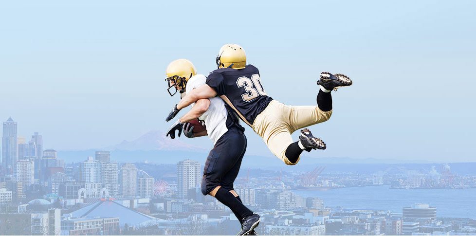
Anterior shoulder instability is the most common form and typically occurs after a traumatic dislocation - often during contact sports or falls. In many cases, the first dislocation results in tearing of the anterior labrum, a fibrocartilaginous rim that deepens the socket and provides stability. This specific tear is called a Bankart lesion and is a leading cause of recurrent instability if not addressed.
Patients often describe symptoms like:
- A sense of the shoulder "slipping" or "giving out"
- Pain or apprehension during certain movements (especially abduction and external rotation)
- Decreased shoulder performance in athletic activities
- Repeated episodes of dislocation or subluxation
Over time, recurrent instability can lead to further damage, including cartilage injury and bone loss, making early and accurate diagnosis critical.
Associated Bony Pathologies: Beyond the Labrum
While soft tissue injury is the hallmark of anterior instability, associated bony injuries are common and significantly impact treatment planning.
- Hill-Sachs Lesion: A compression fracture or dent in the posterior-lateral aspect of the humeral head caused by impact with the anterior glenoid during dislocation. Larger or "engaging" Hill-Sachs lesions can predispose patients to recurrent dislocations.
- Bony Bankart Lesion: In some cases, a piece of the glenoid bone is avulsed along with the labrum, leading to a bony defect in the anteroinferior glenoid rim.
- Glenoid Bone Loss: With recurrent dislocations or prior failed surgeries, patients may develop significant attritional bone loss, often unnoticed without advanced imaging.
These pathologies can alter the biomechanics of the shoulder, reducing the concavity-compression effect and making soft tissue repair alone insufficient in some cases.
Comprehensive Imaging: The Key to Successful Surgical Planning
Accurate imaging is essential in identifying the full extent of shoulder instability and any associated pathology. A thorough workup often includes:
1. Plain Radiographs
Standard X-rays are used to identify fractures, assess joint alignment, and screen for significant Hill-Sachs or bony Bankart lesions. Key views include:
- AP (internal and external rotation)
- Axillary lateral
- Stryker notch view (for Hill-Sachs lesions)
- West Point view (for glenoid rim evaluation)
2. MRI or MR Arthrogram
MRI is the gold standard for evaluating soft tissue injury, including labral tears, capsular laxity, and rotator cuff integrity. An MR arthrogram, which involves the injection of contrast into the joint, provides superior detail for detecting labral pathology and subtle tears.
3. CT Scan with 3D Reconstruction
CT imaging, particularly with 3D reconstructions, is vital in quantifying glenoid bone loss and characterizing bony Bankart and Hill-Sachs lesions. This modality allows for precise measurement of bone deficits, which can guide the decision to perform bone augmentation procedures.
In complex cases, advanced software-assisted 3D modeling can help simulate shoulder movement and predict engagement of Hill-Sachs lesions, guiding preoperative strategy.
When Is Surgery Indicated?
While some patients respond well to physical therapy and activity modification, particularly after a first-time dislocation, surgery is often required in the following scenarios:
- Recurrent shoulder dislocations or subluxations
- High-demand athletes at risk of recurrence
- Large Bankart or Hill-Sachs lesions
- Failure of conservative treatment
- Presence of glenoid bone loss or bony instability
Early surgical intervention in these populations has been shown to improve long-term outcomes and reduce the risk of arthritis associated with chronic instability.
Surgical Treatment Options: Tailored to the Pathology
At our practice, we approach shoulder instability surgery with a patient-specific strategy. Treatment depends on the severity of instability, degree of bone loss, patient activity level, and prior interventions.
1. Arthroscopic Bankart Repair
This minimally invasive approach is ideal for soft tissue injuries without significant bone loss.
- Anchors are inserted into the glenoid to reattach the torn anterior labrum.
- The capsule is plicated or tightened to restore normal joint mechanics.
- Return to play is typically 4–6 months, depending on the sport.
This procedure boasts a high success rate in properly selected patients, particularly those undergoing early repair after injury.
2. Remplissage Procedure
For patients with engaging Hill-Sachs lesions, this technique involves:
- Arthroscopically "filling" the defect with the infraspinatus tendon and posterior capsule.
- Prevents the humeral head from engaging the glenoid rim during movement.
Remplissage is often combined with Bankart repair to reduce the risk of recurrence in patients with bipolar bone defects.
3. Bone Augmentation Procedures
Patients with significant glenoid bone loss (>15–20%) or prior failed stabilization may require more complex solutions:
- Latarjet Procedure: The coracoid process is transferred to the anterior glenoid to increase bone surface and reinforce the anterior capsule with the conjoint tendon. It provides a "triple effect" of bony support, sling effect, and capsular repair.
- Distal Tibial Allograft: Used in select revision cases or where native anatomy limits use of the coracoid. Offers an anatomical contour and cartilage surface for glenoid reconstruction.
These techniques are reserved for high-demand or complex instability and are associated with robust return-to-sport outcomes when executed properly.
Postoperative Recovery & Rehabilitation
Recovery from shoulder stabilization surgery requires dedication and a structured rehab protocol:
Phase 1 (0–4 weeks):
- Sling immobilization to protect the repair
- Passive range of motion initiated under guidance
Phase 2 (4–8 weeks):
- Gradual introduction of active range of motion
- Begin light isometric strengthening
Phase 3 (8–16 weeks):
- Progress to full motion and resistive strengthening
- Proprioceptive and neuromuscular training
Phase 4 (4–6 months):
- Return to sport-specific activities
- Full return to contact sports typically allowed around 6 months
Outcomes are generally excellent, with over 85–90% of patients returning to full activity and sport. Factors like young age, contact sports, and untreated bone loss are associated with higher recurrence.
What If the First Surgery Doesn’t Work?
While outcomes for primary repair are excellent, a small percentage of patients may experience recurrent instability. Common causes include:
- Unrecognized or untreated bone loss
- Traumatic reinjury
- Capsular laxity or poor healing
In these cases, revision stabilization surgery is often successful. Options include:
- Arthroscopic revision with capsular shift
- Open Bankart repair
- Bone block procedures like Latarjet or Eden-Hybinette
- Capsular reconstruction using allograft tissue in cases of severe deficiency
A careful workup - including repeat MRI and CT scans - is essential to identify the cause of failure and ensure long-term success with the next procedure.
Conclusion: Experience Matters in Shoulder Stabilization
Shoulder instability and anterior labral injuries are complex conditions that require a comprehensive understanding of shoulder anatomy, biomechanics, and patient-specific factors. At our practice, we combine high-resolution imaging, advanced arthroscopic techniques, and open surgical strategies to restore shoulder stability and performance.
Whether you’re a high school athlete recovering from your first dislocation or an adult with chronic instability, we’re here to guide you through diagnosis, treatment, and recovery - back to the activities you love.


